Login to your account
Change password, your password must have 8 characters or more and contain 3 of the following:.
- a lower case character,
- an upper case character,
- a special character

Password Changed Successfully
Your password has been changed
Create a new account
Can't sign in? Forgot your password?
Enter your email address below and we will send you the reset instructions
If the address matches an existing account you will receive an email with instructions to reset your password
Request Username
Can't sign in? Forgot your username?
Enter your email address below and we will send you your username
If the address matches an existing account you will receive an email with instructions to retrieve your username

- Institutional Access
Cookies Notification
Our site uses Javascript to enchance its usability. You can disable your ad blocker or whitelist our website www.worldscientific.com to view the full content.
Select your blocker:, adblock plus instructions.
- Click the AdBlock Plus icon in the extension bar
- Click the blue power button
- Click refresh
Adblock Instructions
- Click the AdBlock icon
- Click "Don't run on pages on this site"
uBlock Origin Instructions
- Click on the uBlock Origin icon in the extension bar
- Click on the big, blue power button
- Refresh the web page
uBlock Instructions
- Click on the uBlock icon in the extension bar
Adguard Instructions
- Click on the Adguard icon in the extension bar
- Click on the toggle next to the "Protection on this website" text
Brave Instructions
- Click on the orange lion icon to the right of the address bar
- Click the toggle on the top right, shifting from "Up" to "Down
Adremover Instructions
- Click on the AdRemover icon in the extension bar
- Click the "Don’t run on pages on this domain" button
- Click "Exclude"
Adblock Genesis Instructions
- Click on the Adblock Genesis icon in the extension bar
- Click on the button that says "Whitelist Website"
Super Adblocker Instructions
- Click on the Super Adblocker icon in the extension bar
- Click on the "Don’t run on pages on this domain" button
- Click the "Exclude" button on the pop-up
Ultrablock Instructions
- Click on the UltraBlock icon in the extension bar
- Click on the "Disable UltraBlock for ‘domain name here’" button
Ad Aware Instructions
- Click on the AdAware icon in the extension bar
- Click on the large orange power button
Ghostery Instructions
- Click on the Ghostery icon in the extension bar
- Click on the "Trust Site" button
Firefox Tracking Protection Instructions
- Click on the shield icon on the left side of the address bar
- Click on the toggle that says "Enhanced Tracking protection is ON for this site"
Duck Duck Go Instructions
- Click on the DuckDuckGo icon in the extension bar
- Click on the toggle next to the words "Site Privacy Protection"
Privacy Badger Instructions
- Click on the Privacy Badger icon in the extension bar
- Click on the button that says "Disable Privacy Badger for this site"
Disconnect Instructions
- Click on the Disconnect icon in the extension bar
- Click the button that says "Whitelist Site"
Opera Instructions
- Click on the blue shield icon on the right side of the address bar
- Click the toggle next to "Ads are blocked on this site"
System Upgrade on Tue, May 28th, 2024 at 2am (EDT)
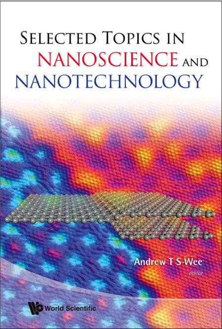
Selected Topics in Nanoscience and Nanotechnology
- Edited by:
- Andrew T S Wee ( NUS, Singapore )
- Add to favorites
- Download Citations
- Track Citations
- Recommend to Library
- Description
- Supplementary
Selected Topics in Nanoscience and Nanotechnology contains a collection of papers in the subfields of scanning probe microscopy, nanofabrication, functional nanoparticles and nanomaterials, molecular engineering and bionanotechnology. Written by experts in their respective fields, it is intended for a general scientific readership who may be non-specialists in these subjects, but who want a reasonably comprehensive introduction to them. This volume is also suitable as resource material for a senior undergraduate or introductory graduate course in nanoscience and nanotechnology.
The review articles have been published in journal COSMOS Vol 3 & 4.
Sample Chapter(s) Chapter 1: Scanning Probe Microscopy Based Nanoscale Patterning and Fabrication (861 KB)
- Scanning Probe Microscopy Based Nanoscale Patterning and Fabrication (X N Xie et al.)
- Nanoscale Characterization by Scanning Tunneling Microscopy (H Xu et al.)
- EUV Lithography for Semiconductor Manufacturing and Nanofabrication (H Kinoshita et al.)
- Synchrotron-Radiation-Supported High-Aspect-Ratio Nanofabrication (A Chen et al.)
- Chemical Interactions at Noble Metal Nanoparticle Surfaces — Catalysis, Sensors and Devices (A S Nair et al.)
- Diamond-Like Carbon: A New Material Base for Nanoarchitectures (X Li & D H C Chua)
- Hotplate Technique for Nanomaterials (Y Zhu & C H Sow)
- π- d Interaction Based Molecular Conducting Magnets: How to Increase the Effects of the π- d Interaction (A Miyazaki & T Enoki)
- Recent Developments on Porphyrin Assemblies (R Charveta et al.)
- Nanostructures from Designer Peptides (B T Ong et al.)
- Nanotechnology and Human Diseases (G Y H Lee & C T Lim)
- Nanomedicine: Nanoparticles of Biodegradable Polymers for Cancer Diagnosis and Treatment (S S Feng)
FRONT MATTER
- Andrew T. S. Wee
- Pages: i–viii
https://doi.org/10.1142/9789812839565_fmatter
Scanning Probe Techniques
Scanning probe microscopy based nanoscale patterning and fabrication.
- XIAN NING XIE ,
- HONG JING CHUNG , and
- ANDREW THYE SHEN WEE
- Pages: 3–23
https://doi.org/10.1142/9789812839565_0001
Nanotechnology is vital to the fabrication of integrated circuits, memory devices, display units, biochips and biosensors. Scanning probe microscope (SPM) has emerged to be a unique tool for materials structuring and patterning with atomic and molecular resolution. SPM includes scanning tunneling microscopy (STM) and atomic force microscopy (AFM). In this chapter, we selectively discuss the atomic and molecular manipulation capabilities of STM nanolithography. As for AFM nanolithography, we focus on those nanopatterning techniques involving water and/or air when operated in ambient. The typical methods, mechanisms and applications of selected SPM nanolithographic techniques in nanoscale structuring and fabrication are reviewed.
NANOSCALE CHARACTERIZATION BY SCANNING TUNNELING MICROSCOPY
- M. A. K. ZILANI ,
- WEI CHEN , and
- Pages: 25–52
https://doi.org/10.1142/9789812839565_0002
Nanoscale characterization is a key field in nanoscience and technology as it provides fundamental understanding of the properties and functionalities of materials down to the atomic and molecular scale. In this article, we review the development and application of scanning tunneling microscope (STM) techniques in nanoscale characterization. We will discuss the working principle, experimental setup, operational modes, and tip preparation methods of scanning tunneling microscope. Selected examples are provided to illustrate the application of STM in the nanocharacterization of semiconductors. In addition, new developments in STM techniques including spin-polarized STM (SP-STM) and multiprobe STM (MP-STM) are discussed in comparison with conventional non-magnetic and single tip STM methods.
Nanofabrication
Euv lithography for semiconductor manufacturing and nanofabrication.
- HIROO KINOSHITA
- Pages: 55–81
https://doi.org/10.1142/9789812839565_0003
EUV lithography is the exposure technology in which even 15 nm node which is the limit of Si device can be achieved. Unlike the conventional optical lithography, this technology serves as a reflection type optical system, and a multilayer coated mirror is used. Development of manufacturing equipment is accelerated to aim at the utilization starting from 2011. The critical issues of development are the EUV light source which has the power over 115 W and resist with high sensitivity and low line edge roughness (LER).
SYNCHROTRON-RADIATION-SUPPORTED HIGH-ASPECT-RATIO NANOFABRICATION
- L. K. JIAN , and
- HERBERT O. MOSER
- Pages: 83–92
https://doi.org/10.1142/9789812839565_0004
X-ray lithography with synchrotron radiation is an important nanolithographic tool which has unique advantages in the production of high aspect ratio nanostructures. The optimum synchrotron radiation spectrum for nanometer scale X-ray lithography is normally in the range of 500 eV to 2 keV. In this paper, we present the main methods, equipment, process parameters and preliminary results of nanofabrication by proximity X-ray lithography within the nanomanufacturing program pursued by Singapore Synchrotron Light Source (SSLS). Nanostructures with feature sizes down to 200 nm and an aspect ratio up to 10 have been successfully achieved by this approach.
Functional Nanomaterials
Chemical interactions at noble metal nanoparticle surfaces — catalysis, sensors and devices.
- A. SREEKUMARAN NAIR ,
- RENJIS T. TOM ,
- V. R. RAJEEV KUMAR ,
- C. SUBRAMANIAM , and
- Pages: 95–116
https://doi.org/10.1142/9789812839565_0005
In this paper, a summary of some of the recent research efforts in our laboratory on chemical interactions at noble metal nanoparticle surfaces is presented. The article is divided into five sections, detailing with (i) interactions of simple halocarbons with gold and silver nanoparticle surfaces at room temperature by a new chemistry and the exploitation of this chemistry in the extraction of pesticides from drinking water, (ii) interaction of biologically important proteins such as Cyt c , hemoglobin and myoglobin as well as a model system, hemin with gold and silver nanoparticles and nanorods forming nano–bio conjugates and their surface binding chemistry, (iii) formation of polymer–nano composites with tunable optical properties and temperature sensing characteristics by single and multi-step methodologies, (iv) nanomaterials-based flow sensors and (v) composites of noble metal nanoparticles and metallic carbon nanotubes showing visible fluorescence induced by metal–semiconductor transition.
DIAMOND-LIKE CARBON: A NEW MATERIAL BASE FOR NANO-ARCHITECTURES
- XIJUN LI and
- DANIEL H. C. CHUA
- Pages: 117–148
https://doi.org/10.1142/9789812839565_0006
Diamond-like carbon (DLC) is a form of amorphous carbon which has high fraction of sp 3 hybridization. Due to its nature of sp 3 bonding, diamond-like carbon has been shown to have excellent properties similar to that of diamond. This includes high hardness, excellent wear-resistance, large modulus and chemically inert. Traditional applications include wear resistant coatings and protective film. This article intends to review the synthesis and material properties of diamond-like carbon as well as its potential as a novel material for applications in nano-architecture and nano-mechanical devices. An introduction into metal-dopants in diamond-like carbon film will be briefly mentioned as well as techniques on the design and fabrication of this material.
HOTPLATE TECHNIQUE FOR NANOMATERIALS
- YANWU ZHU and
- CHORNG HAUR SOW
- Pages: 149–169
https://doi.org/10.1142/9789812839565_0007
As an efficient and cost-effective method to synthesize nanomaterials, the hotplate technique has been reviewed in this article. Systematic studies have been carried out on the characterizations of the materials synthesized. In addition to the direct preparation of nanomaterials on metals, this method has been extended to the substrate-friendly and plasma-assisted hotplate synthesis. Apart from chemically pure nanostructures, a few nanohybrids were synthesized, further demonstrating the flexibility of this technique. The investigations on their applications indicate that they are promising material systems with potential applications in field emission devices, gas sensors, Li-ion batteries and ultrafast optical devices.
Molecular Engineering
Π–d interaction based molecular conducting magnets: how to increase the effects of the π–d interaction.
- AKIRA MIYAZAKI and
- TOSHIAKI ENOKI
- Pages: 173–182
https://doi.org/10.1142/9789812839565_0008
The crystal structures and electronic and magnetic properties of conducting molecular magnets developed by our group are reviewed from the viewpoints of our two current strategies for increasing the efficiency of the π–d interaction. (EDTDM) 2 FeBr 4 is composed of quasi-one-dimensional donor sheets sandwiched between magnetic anion sheets. The ground state of the donor layer changes from the insulator state to the metallic state by the application of pressure. When it is near to the insulator-metal phase boundary pressure, the magnetic order of the anion spins considerably affects the transport properties of the donor layer. The crystal structure of (EDO–TTFBr 2 ) 2 FeX 4 (X = Cl, Br) is characterized by strong intermolecular halogen-halogen contacts between the organic donor and FeX 4 anion molecules. The presence of the magnetic order of the Fe 3+ spins and relatively high magnetic order transition temperature proves the role of the halogen-halogen contacts as exchange interaction paths.
RECENT DEVELOPMENTS ON PORPHYRIN ASSEMBLIES
- RICHARD CHARVET ,
- JONATHAN P. HILL ,
- YONGSHU XIE ,
- YUTAKA WAKAYAMA , and
- KATSUHIKO ARIGA
- Pages: 183–213
https://doi.org/10.1142/9789812839565_0009
The porphyrin macrocycle is one of the most frequently investigated functional molecular entities and can be incorporated into advanced functional nanomaterials upon formation of organized nanostructures. Thus, study of the science and technology of porphyrin assemblies has attracted many organic, biological and supramolecular chemists. A wide variety of nanostructures can be obtained by supramolecular self-assembly because the porphyrin moiety is amenable to chemical modifications through thoughtful synthetic design and moderate preparative effort. Some recent developments in porphyrin assembly, obtained through various supramolecular approaches, are briefly summarized. Topics described in this review are classified into four categories: (i) non-specific assemblies; (ii) specific assemblies; (iii) assemblies in organized films; (iv) molecular-level arrangement. We present examples in the order of structural precision of assemblies.
Bionanotechnology and Nanomedicine
Nanostructures from designer peptides.
- BOON TEE ONG ,
- PARAYIL KUMARAN AJIKUMAR , and
- SURESH VALIYAVEETTIL
- Pages: 217–227
https://doi.org/10.1142/9789812839565_0010
The present article reviews the self-assembly of oligopeptides to form nanostructures, both in solution and in solid state. The solution structures of the peptides were examined using circular dichroism and dynamic light scattering. The solid state assembly was examined by adsorbing the peptides onto a mica surface and analyzing it using atomic force microscopy. The role of pH and salt concentration on the peptide self-assembly was also examined. Nanostructures within a size range of 3–10 nm were obtained under different conditions.
NANOTECHNOLOGY AND HUMAN DISEASES
- GABRIEL YEW HOE LEE and
- CHWEE TECK LIM
- Pages: 229–241
https://doi.org/10.1142/9789812839565_0011
Tissues, cells and biomolecules can experience changes in their structural and mechanical properties during the occurrence of certain diseases. Recent advances in the fields of nanotechnology, biomechanics and cell and molecular biology have led to the development of state-of-the-art and novel biophysical and nanotechnological tools to probe the mechanical properties of individual living cells and biomolecules. Here we will review the basic principles and application of some of these nanotechnological tools used to relate changes in the elastic and viscoelastic properties of cells to alterations in the cellular and molecular structures induced by diseases such as malaria and cancer. Knowing the ways and the extent to which mechanical properties of living cells are altered with the onset of disease progression will be crucial for us to gain vital insights into the pathogenesis and pathophysiology of malaria and cancer, and potentially offers the opportunity to develop new and better methods of detection, diagnosis and treatment.
NANOMEDICINE: NANOPARTICLES OF BIODEGRADABLE POLYMERS FOR CANCER DIAGNOSIS AND TREATMENT
- Pages: 243–259
https://doi.org/10.1142/9789812839565_0012
Nanomedicine is to apply and further develop nanotechnology to solve problems in medicine, i.e. to diagnose, treat and prevent diseases at the cellular and molecular level. This article demonstrates through a full spectrum of proof-of-concept research, from nanoparticle preparation and characterization, in vitro drug release and cytotoxicity, to in vivo pharmacokinetics and xenograft model, how nanoparticles of biodegradable polymers could provide an ideal solution for the problems encountered in the current regimen of chemotherapy. A system of vitamin E TPGS coated poly(lactic-co-glycolic acid) (PLGA) nanoparticles is used as an example for paclitaxel formulation as a model drug. In vitro HT-29 cancer cell viability experiment demonstrated that the paclitaxel formulated in the nanoparticles could be 5.64 times more effective than Taxol ® after 24 hr of treatment. In vivo pharmacokinetics showed that the drug formulated in the nanoparticles could achieve 3.9 times higher therapeutic effects judged by area-under-the curve (AUC). One shot can realize sustainable chemotherapy of 168 hr compared with 22 hr for Taxol ® at a single 10mg/kg dose. Xenograft tumor model further confirmed the advantages of the nanoparticle formulation versus Taxol ® .
Sample Chapter(s) Chapter 1: Scanning Probe Microscopy Based Nanoscale Patterning and Fabrication (861k)

Related Books

Nano-Engineering in Science and Technology

Silicon Carbide Microelectromechanical Systems for Harsh Environments

Theory of Semiconductor Quantum Dots

Current at the Nanoscale

Embedded Dielectrics for Electronic Packaging

Principles of Nanotechnology

Emerging Nanotechnology Power

Nanosciences

Advances in Multiphysics Simulation and Experimental Testing of Mems

Annual Review of Nano Research

Handbook of Nanobiomedical Research

Nanobioceramics for Healthcare Applications

Nanotechnology Challenges

Asia Pacific Nanotechnology Forum 2003

Diamondoid Molecules

Size Really Does Matter

Static and Dynamic Problems of Nanobeams and Nanoplates

Nanotechnology from lab to industry – a look at current trends

First published on 1st August 2022
Nanotechnology holds great promise and is hyped by many as the next industrial evolution. Medicine, food and cosmetics, agriculture and environmental health, and technology industries already profit from nanotechnology innovations and their influence is expected to increase drastically in the near future. However, there are also many challenges that need to be overcome to bring a nanotechnological product or business to the market. In this article we discuss current examples of nanotechnology that have been successfully introduced in the market and their relevance and geographical spread. We then discuss different partners for scientists and their role in the commercialization process. Finally, we review the different steps it takes to bring a nanotechnology to the market, highlight the many difficulties related to these steps, and provide a roadmap for the journey from lab to industry which can be beneficial to researchers.

1. Introduction
| Possible applications of nanomaterials. | ||
2. Nanotechnology developments
| The top 25 countries involved in the publishing of nanotechnology discoveries. (a) And patenting of inventions including at least one claim related to nanotechnology or patents classified with an International Patent Classification (IPC) code related to nanotechnology in the year 2020. (b) (https://statnano.com/). | ||
All inhabited continents are represented among the top countries involved in scientific publishing; however, only Europe (14 countries), Asia (8 countries), North America (2 countries), and Oceania (1 country) are included among the top 25 countries involved in the patenting of nanotechnology developments. Seventeen countries were common factors among both publishing and patenting discoveries. It is also noteworthy that the two countries that had the highest investments in scientific research (China and The USA) produced highest numbers of publications and patents, respectively. Patents can be used as technological indicators as they provide an insight into the research and development activities that are intended for commercial gain. 12 The transfer of these nanotechnology advancements to commercialized end products is however a major challenge that the scientific community faces. However, it has to be noted here that there are also quite some differences in culture when it comes to patenting. There are differences between countries in how buerocratic the patent process is. Additionally, there are differences in how much is patented at all. In some cultures, it might be more common to keep innovation a secret than to patent. There are also differences in how patents are made. In some places there is a high number of smaller patents while in others there are a few more elaborate ones.
2.1. Nanotechnology industries worldwide
| Company | Operation | Country |
|---|---|---|
| Operations listed might not be exhaustive. | ||
| 3M | Manufactures numerous nanomaterials | USA |
| Advanced Material Development | Develops 2D nanotechnologies and metamaterial systems | UK |
| Applied Graphene Materials | Develops and applies graphene nanoplatelet dispersions | UK |
| BNNano, Inc. | Manufactures boron nitride nanotubes (NanoBarbs™) | USA |
| CelluForce | Produces a form of cellulose nanocrystals (CelluForce NCC™) | Canada |
| Cerion | Manufactures metal, metal oxide, and ceramic nanomaterials | USA |
| INNOVNANO | Manufactures ultra-fine nanostructured ceramic powders | Portugal |
| Nanogap | Manufactures novel nanomaterials from atomic quantum clusters | Spain |
| Nanomakers | Develops and commercializes nanoparticles of silicon carbide | France |
| OCSiAl Luxembourg | Produces graphene nanotubes | Luxembourg |
| RAS AG | Produces and distributes of nanomaterials | Germany |
| Rezenerate NanoFacial | Develops nanofacials using innovative devices for cosmetics delivery | USA |
| Superbranche | Develops functionalized metallic oxide nanoparticles | France |
| Zeon Corporation | Manufactures single-walled carbon nanotube | Japan |
3. The business of ‘lab-to-industry’
| Roadmap for the commercialization of nanotechnology-derived products. | ||
3.1. Ideation
These two approaches show how innovation relies on technology seeds and market needs. One might ponder which of the two approaches is better. There are both merits and challenges associated with each approach. While each can lead to innovation, a pairing of the two is recommended. When closely integrated, the potential impact of the innovation increases. This synchronization of the ‘seed’ and ‘need’ approaches is called accelerated innovation. It enables the restructuring of research and development, and innovation processes to make new product development dramatically faster and less costly. 15 Furthermore, it also facilitates functional thinking and exaptation where the latter refers to the discovery of unintended functions for technologies. Altogether creating the ideal conditions for researchers to make radical innovations and bridge the gap between academia and industry.
3.2. Business model
Breakthrough technologies, especially those incorporating the use of nanotechnology, are intended to create value. Value is created via this technology when there is meaningful performance improvement or when the cost of solving problems is significantly reduced. There is however a major challenge for nanotechnology innovations in terms of a business model, and that is, the challenge of taking the product to customers. Several factors can influence this (for example, having limited resources) and for this reason, a go-to-market strategy is critical.
A joint-development partnership is an agreement between two organizations to develop a new product or service. It is a strategic alliance that serves to leverage the assets of each company to create a new offering for commercialization that would be difficult to achieve individually. This type of partnership is commonly used for product development or beta testing. Typically, these agreements are not binding and one party can quit at any time. Profits, access, expenses, and losses are usually shared between the companies. With this type of business partnership, it is important to have a close business relationship with the company before engaging in this agreement. As is the case with licensing arrangements, the most ideal joint-development partnership can be determined with the assistance of an attorney. Matters relating to the ownership and access to intellectual property, responsibilities, disengagement, and termination are some of the issues to be discussed with a suitable attorney before engaging a potential partner.
In partnerships, securing intellectual property early remains crucial. In an innovative nanotechnology business, the science underpinning the technology is critical and must be protected. This can be achieved by engaging an intellectual property counsel. The services of a corporate counsel should also be acquired early to ensure the start-up is properly incorporated. These parties should be appointed at the early stages as they help with structuring the company. The technology transfer process which is discussed in Section 3.3 helps to get these counsels on board.
There are some key players that are needed to guarantee a good business model and these are outlined in Fig. 4 . To assure a diversity of skills that are necessary for success, an often overlooked group of individuals is needed. This is a company board. This can include a board of advisors and a board of directors. The functions of these two bodies bear some similarities and differences. The board of advisors is composed of business professionals who fill skill and expertise gaps and can offer guidance to the management team. This can include matters concerning business performance, market trends, long-term goals of the company, and financing to name a few. While the additional skill set required in a science-based industry might be in business management, it is not unusual for additional technical expertise to be warranted. This can include the skills of fellow scientists who have had prior success in transitioning science to the marketplace. These scientists, when recruited, could form a scientific or technical advisory board. Regardless of the composition of the advisory board, their core function is to provide non-binding strategic advice. Their role is not fiduciary. This means that the team of experts and community leaders has no legal responsibility to the company. Their role however remains critical as they can compensate for some of the weaknesses within the management team and bring different opinions, perspectives, and experiences to the table. The board of advisors is particularly helpful for start-ups. A board of directors, on the other hand, is essentially a panel of people elected or appointed to represent shareholders. They oversee the activities of the company and have a fiduciary responsibility to represent and protect the members' or investors' interests in the company. The management team however reports to the board of directors. Larger companies that will require significant funding need a board of directors. Both the boards of advisors and directors can assist with strategic planning, the development of new ideas, improvement of management structure, improving company image and reputation, reassuring stakeholders and investors, and overall, help to ensure the success of the company.
| Key players to support a budding nanotechnology start-up. | ||
The management team and the company board can together decide on the most suitable business model for the company. In making this decision, special focus should be placed on the model that will create and deliver great value to customers while simultaneously delivering great margins. The model should also hedge against customer dissatisfaction or dissonance and issues securing adequate funding. While the team is now multifaceted, additional support to make the right decisions that will position the company for success can be sought. This can be achieved using accelerators and incubators (which might be available within the university or municipality), government agencies such as the local chamber of commerce, and small business and technology development centers. Start-ups are generally encouraged to not employ at the early stages and to instead contract personnel for specific functions if necessary.
3.3. Technology transfer process
The efficiency of the transfer of nanotechnology innovations from the lab to the industry is dependent on the efficacy of the technology transfer process. Countries that invest in improving nanotechnology transfer policies and practices have greater nanotechnology outputs. This is evident in the United States where the National Nanotechnology Initiative (NNI) was developed. It is a collaboration of federal departments and agencies with interests in nanotechnology research, development, and commercialization. 17 Within the NNI are agencies such as the Nano manufacturing and Small Business Innovation Research (SBIR) programs, and the NNI's National Nanotechnology Coordination Office (NNCO) that are concerned with the transfer of newly developed nanotechnologies into products for commercial use. In Asia, there has been an increase in expenditure towards nanotechnology research and deliberate efforts to transfer research findings to industries. While the production of nanotechnology publications in China is higher than in other countries ( Fig. 2a ), the transfer of these technologies to industries is not equivalent. 18 The National Steering Committee for Nanoscience and Nanotechnology (NSCNN) was established to oversee and coordinate nanotechnology policies and programs in China. Some key members of this group include the Chinese Academy of Sciences (CAS), the National Natural Science Foundation of China (NSFC), the National Development and Reform Commission (NDRC), and the Chinese Academy of Engineering. These agencies are expected to impact the technology transfer process within the country.
The success of the transfer of technology in The United States reveals that more favorable environments for nanotechnology transfer need to be created globally. This will create a stronger ecosystem for nanotechnology research and innovation, and in turn, result in greater success in the use of intellectual property to facilitate the creation of start-ups formed from the ground up or through partnerships. Some nanotechnology and nano-engineering associations across the world that can be modelled in other countries to positively impact the transfer of technology are outlined in Table 2 . These associations were selected from the Nanotechnology 2020 Market Analysis. 9
| Association | Country |
|---|---|
| Alliance for Nanotechnology in Cancer | USA |
| American National Standards Institute Nanotechnology Panel | USA |
| Centre for Nano and Soft Matter Sciences | India |
| Collaborative Centre for Applied Nanotechnology | Ireland |
| Indian Association for the Cultivation of Science | India |
| Iranian Nanotechnology Laboratory Network | Iran |
| Nano Medicine Roadmap Initiative | USA |
| National Cancer Institute | USA |
| National Institutes of Health | USA |
| National Research Council Nanotechnology Research Centre | Canada |
| Russian Nanotechnology Corporation | Russia |
| S.N. Bose national Centre for Basic Sciences | India |
| Waterloo Institute for Nanotechnology | Canada |
3.4. Readiness for commercialization
| A cloverleaf framework for market entry readiness assessment of nanotechnology inventions. | ||
Technology readiness evaluates the technology itself and seeks to determine if the technology will maintain itself in the market. This is usually determined by performing a technology readiness assessment (TRA). It is recommended that this TRA is done at several points during the ‘life cycle’ of the new technology or system. Possible components of this assessment include an evaluation of the conceptual design, a clear protocol to facilitate a decision from among several competing design options, and similarly, a defined approach to decide when to begin full-scale development. These decisions might be made by the research team or they can be more complex and warrant an external, independent peer-review process. 20 Market readiness assesses how marketable the technology is; that is, how well the technology will be accepted by the target market. This is generally done by examining whether the technology offers meaningful identifiable and quantifiable benefits, has distinct advantages over competing products, has access to a market of a suitable size that is defined and is growing (demand-based), has immediate market uses, and has feasible manufacturing requirements. 21
The commercialization readiness assessment also evaluates the readiness of the technology's business model. This is done to verify the stability and readiness of the foundation upon which the technology will be delivered. Within this component, parameters for assessment include determining whether prospective licensees are identified, if industry contacts are available, and if further development or patenting is possible based on the availability of financial support for the licensee. Additionally, anticipated future royalty revenue of the license, access to venture capital, a profitable investment, and availability of government support for additional development for innovations resulting from universities are also crucial. 22 The last key area is management readiness which assesses the readiness of the management team that is responsible for the technology. It addresses matters such as the ability of the inventor to champion the innovation as a team player, whether the inventor's expectations for success are realistic, if the inventor is recognized and reputable in the field, if commercialization skills such as sales and marketing skills are available, whether management capabilities are available, and also whether the inventor is the patent holder for innovations resulting from government labs. 23
A method of quantifying the judgments made for each criterion of the four areas of the Cloverleaf framework to determine the degree to which each condition is met was suggested. 19 If all components of the criteria list for the four ‘leaves’ assessing readiness are satisfied, then the technology is ready for commercialization. If a partnership agreement is being utilized, some components should be completed before engaging a partner and others should be finalized with the partner. Regardless of the business model, if any area is found lacking, additional preparation is warranted to ensure the success of the venture when it enters the market.
Alternative to the Cloverleaf framework is the Technology Readiness Levels (TRL) model. This was developed by NASA and is a type of measurement system that is used to permit more effective assessment and communication regarding the maturity of new technologies. 20 The different levels of the framework are outlined in Fig. 6 . There are nine technology readiness levels. A project is evaluated against the parameters for each technology level and is then assigned a TRL rating based on its progress. TRL 1 is the lowest level and indicates that a technology requires further research and development, and testing. TRL 9 is the highest level and signifies a mature technology that is proven to work and may be put into use and commercialized.
| Technology readiness levels (TRL). | ||
3.5. Financials
| Phases of a company's growth (a), (b) and the different funding instruments that are available at the different stages (c). | ||
Another type of capital provider is venture capitalists. These private investors provide funds to early-stage companies that are pursuing big opportunities with high growth potential. Venture capital firms exchange capital for equity ownership and can also provide strategic assistance, and an invaluable network. To capture the interest of a venture capitalist, a start-up should have a good “elevator pitch” and a strong investor pitch deck for their innovative product. This should therefore include the strength of the management team and clearly outline the large potential market for the nanotechnology innovation, and a unique product or service with a strong competitive advantage. Another entity that can provide financing and has a similar structure to a venture capital firm is a family office. This is a special investment firm that manages the wealth owned by individuals and families with a high net worth. 26 Family offices make optimal investors and are increasingly entering venture investment as a relatively new capital provider. They are comprised of qualified professionals with extensive experience and tend to offer more patient capital and expect lower returns than traditional investors.
4. The challenge of moving technology from lab to industry
| Summary of start-up lifetime and the most common reasons for failure. Adapted from Cantamessa et al. with permissions from MDPI. | ||
Biological or environmental challenges are other factors that can impede the transfer of nanotechnology from the lab to the industry. Biological challenges include insufficient knowledge involving the interaction of nanomaterials in vitro and in vivo , inadequate information on their bioaccumulation in target organs, tissues, and cells, and also limited information on their biocompatibility. 30,31 Physical properties such as particle size, composition, surface area, surface charge, surface chemistry, and agglomeration state all influence the biocompatibility of nanomaterials and so more information is needed on their safety in vivo . 31 Environmental challenges include nanomaterials entering the environment either directly or indirectly (for example, via landfills). Nanomaterials can have potentially adverse effects on natural systems and can enter the environment at different stages of their life cycle. Three emission scenarios that are generally of relevance are (i) release during the production of various nanotechnology products or nano-enabled products; (ii) release during use; and (iii) release after disposal. 32 While present in the environment, nanomaterials can then undergo many transformations. These include chemical transformations (for example, photo-degradation), physical transformations (such as aggregation), biologically-mediated transformations (for instance, redox reactions in biological systems), and interactions with macromolecules (for example, flocculation). 30 The interplay between these transformations and the transport of the nanomaterial within the ecosystem ultimately determine their fate and ecotoxicity.
Possible biological and environmental impacts of nanotechnology innovations should be determined with in vitro and in vivo models, as well as within aquatic and terrestrial ecosystems. The production process from which the nanomaterial results should also be considered so that any such material emitted during this time or released from nano-enabled devices during their fabrication, use, recycling or disposal can be studied and minimized. Biological and environmental challenges can also be mitigated by providing employers and the extended workforce with information on the potential toxicity of nanomaterials at different stages of their life cycle. With the help of modelling, recent developments have been geared towards predicting the fate, behavior, and concentration of nanomaterials in the environment. 33 While these simulations can be helpful, more efficient and reliable analytical instruments and methods must be developed so that nanomaterials can be satisfactorily characterized and quantified, and the necessary tools developed to detect, monitor and track them in biological media and complex environmental matrixes.
The nanotechnology industry plays a major role in economic development; however, several economic challenges can hinder the transfer of innovations from the lab to the industry. Generally, these include limited investment in relevant research and development activities and a lack of appropriate mechanisms to secure these investments, lack of laboratory equipment and appropriate infrastructure to facilitate research and its commercialization, and insufficient funding opportunities to engage in research that has the potential for commercialization. Constraints imposed on the activities needed to commercialize nanotechnology outputs are also impacted by the socio-economic dynamics of innovation. While many believe the rapid growth in nanotechnology will have significant economic benefits, some advocate to reduce or halt its development. The backlash against nanotechnology by this group is based on the belief that it will exacerbate problems concerning existing socio-economic inequity and power imbalance caused by inequality. This, they suggest, will cause a nano-divide which refers to differing access to nanotechnology between low-, middle-, and high-income countries. 34,35 The ethical criticism is mainly concerned with inequity based on where knowledge is developed and retained and a country's capacity to engage in these processes. 35 An attempt to combat these challenges is outlined in the European Union's Framework Programs through the Responsible Research and Innovation (RRI) approach. This approach ‘anticipates and assesses potential implications and societal expectations concerning research and innovation, intending to foster the design of inclusive and sustainable research and innovation’ (https://ec.europa.eu). These measures which are intended to facilitate broader access to nano-technology and its innovations globally are critical in addressing a nano-divide.
The final category of challenges that can significantly impact the transfer of nanotechnology from the lab to the industry is regulatory challenges. These are concerned with a lack of clear regulatory guidelines for nanotechnology and nanotechnology-enabled products. Some regulatory challenges include inadequate policies to foster the development and operation of nanotechnology businesses or insufficient strategies implemented by governments to attract nanotechnology business initiatives. Additionally, a lack of technology transfer protocols, or requisites for regulatory approvals to facilitate the movement of innovation from the lab to commercial products are problematic. 36 The multidisciplinary nature of nanotechnology also presents regulatory challenges. With its cross-industry applications, policing and enforcement nanotechnology patents have proven to be prohibitively expensive (WIPO, 2011). New intellectual property practices and protocols are therefore required to simplify the pathway from lab to industry thereby reducing time and expense.
The technical, biological, environmental, economic, and regulatory challenges of nanotechnology need to be addressed urgently. Policies governing all aspects of nanotechnology research and subsequent commercialization must balance its potential benefits with its current challenges. Combatting these challenges will require considerable efforts to prevent any possible harmful effects of nanotechnology while also facilitating the awareness of its benefits to society. 37 The involvement of scientific, governmental, industry, and labor force representatives is therefore critical in decision making so the challenges associated with the commercialization of nanotechnology can be controlled, minimized or mitigated.
5. Conclusions
The necessary risk assessment to understand the potentially harmful effects of products resulting from nanotechnology have however not kept pace with their proliferation; and researchers are racing to address this knowledge gap. 38 Companies resulting from the transfer of nanotechnology innovations from the lab to the marketplace must therefore have rigorous risk management protocols where risks are identified, control measures are planned and implemented, and risks communication. 37 Identified regulatory impediments should also be addressed and technology transfer policies and practices implemented. Entrepreneurial education and training, and the establishment of business incubators should also be supported within the necessary departments or research institutes. Improvement in the understanding of nanotechnology within society would also help commercialization efforts. Overall, societal actors such as researchers, policymakers, investors, citizens etc. must work together during the research and commercialization stages so that the many benefits of nanotechnology outputs can be aligned with the needs and expectations of society.
Conflicts of interest
Notes and references.
- M. U. Munir, D.-N. Phan and M. Q. Khan, Nanomaterials Recycling , 2022, pp. 209–222 Search PubMed .
- K. T. Kosmowski, Safety and Reliability of Systems and Processes , 2021 Search PubMed .
- M. Nasrollahzadeh, S. M. Sajadi, M. Sajjadi and Z. Issaabadi M. Atarod, Interface Sci. Technol. , 2019, 28 , 113–143 CrossRef CAS .
- L. Nie, A. Nusantara, V. Damle, R. Sharmin, E. Evans, S. Hemelaar, K. Van der Laan, R. Li, F. Perona Martinez, T. Vedelaar, M. Chipaux and R. Schirhagl, Sci. Adv. , 2021, 7 (21), eabf0573 CrossRef CAS PubMed .
- D. Hälg, T. Gisler, Y. Tsaturyan, L. Catalini, U. Grob, M.-D. Krass, M. Héritier, H. Mattiat, A.-K. Thamm and R. Schirhagl, Phys. Rev. Appl. , 2021, 15 (2), L021001 CrossRef .
- A. Munawar, Y. Ong, R. Schirhagl, M. A. Tahir, W. S. Khan and S. Z. Bajwa, RSC Adv. , 2019, 9 (12), 6793–6803 RSC .
- T. F. Rambaran, Appl. Sci. , 2020, 2 (8), 1–26 Search PubMed .
- A. Nanda, S. Nanda, T. A. Nguyen, S. Rajendran and Y. Slimani, Nanocosmetics , 2020, 3–16 Search PubMed .
- O. Adiguzel, Biomater. Med. Appl. , 2020, 3 (1), 1335 Search PubMed .
- H. Dong, Y. Gao, P. J. Sinko, Z. Wu, J. Xu and L. Jia, Nano Today , 2016, 11 (1), 7–12 CrossRef CAS .
- E. Inshakova and A. Inshakova, IOP Conference Series: Materials Science and Engineering , 2020, IOP Publishing, vol. 3, p. 033020 Search PubMed .
- J. R. Saura, D. Ribeiro-Soriano and D. Palacios-Marqués, Int. J. Inf. Manag. , 2004, 102331 Search PubMed .
- T. F. Rambaran and A. Nordström, Food Frontiers , 2021, 2 (2), 140–152 CrossRef CAS .
- L. Zhang, Y. Tang and L. Tong, iScience , 2020, 23 (1), 100810 CrossRef PubMed .
- P. J. Williamson, Glob. Strategy J. , 2016, 6 (3), 197–210 CrossRef .
- S. Cunningham, Drug Discovery Today , 2020, 25 (8), 1291 CrossRef CAS PubMed .
- M. C. Roco, Handbook on nanoscience, engineering and technology , vol. 2, 2007 Search PubMed .
- Y. Gao, B. Jin, W. Shen, P. J. Sinko, X. Xie, H. Zhang and L. Jia, Nanomed. Nanotechnol., Biol. Med. , 2016, 12 (1), 13–19 CrossRef CAS PubMed .
- L. A. Heslop, E. McGregor and M. Griffith, J. Technol. Tran. , 2001, 26 (4), 369–384 CrossRef .
- J. C. Mankins, Acta Astronaut. , 2009, 65 (9), 1216–1223 CrossRef .
- G. A. Buchner, K. J. Stepputat, A. W. Zimmermann and R. Schomäcker, Ind. Eng. Chem. Res. , 2019, 58 (17), 6957–6969 CrossRef CAS .
- G. A. Van Norman and R. Eisenkot, JACC Basic Transl. Sci. , 2017, 2 (2), 197–208 CrossRef PubMed .
- R. Oosthuizen and A. J. Buys, S. Afr. J. Ind. Eng. , 2003, 14 (1), 111–124 Search PubMed .
- Successful founding and financing of nanotechnology companies , https://www.nanowerk.com/nanotechnology/investing/funding_nanotechnology_companies_1.php, accessed May 10, 2022 Search PubMed .
- M. Cantamessa, V. Gatteschi, G. Perboli and M. Rosano, Sustainability , 2018, 10 (7), 2346 CrossRef .
- D. Kenyon-Rouvinez and J. E. Park, J. Wealth Manag. , 2020, 22 (4), 8–20 CrossRef .
- L. F. Kampers, E. Asin-Garcia, P. J. Schaap, A. Wagemakers and V. A. M. Dos Santos, Trends Biotechnol. , 2021, 39 (12), 1240–1242 CrossRef CAS PubMed .
- E. Prassler, IEEE Robot. Autom. Mag. , 2016, 23 (3), 11–14 Search PubMed .
- I. P. Kaur, V. Kakkar, P. K. Deol, M. Yadav, M. Singh and I. Sharma, J. Controlled Release , 2014, 193 , 51–62 CrossRef CAS PubMed .
- G. V. Lowry, K. B. Gregory, S. C. Apte and J. R. Lead, Transformations of nanomaterials in the environment , ACS Publications, 2012 Search PubMed .
- Y. Yoshioka, K. Higashisaka and Y. Tsutsumi, Nanomaterials in Pharmacology , Springer, 2016, pp. 185–199 Search PubMed .
- M. Bundschuh, J. Filser, S. Lüderwald, M. S. McKee, G. Metreveli, G. E. Schaumann, R. Schulz and S. Wagner, Environ. Sci. Eur. , 2018, 30 (1), 1–17 CrossRef CAS PubMed .
- R. J. Williams, S. Harrison, V. Keller, J. Kuenen, S. Lofts, A. Praetorius, C. Svendsen, L. C. Vermeulen and J. van Wijnen, Curr. Opin. Environ. Sustain. , 2019, 36 , 105–115 CrossRef .
- G. Miller and G. Scrinis, Nanotechnology and the Challenges of Equity, Equality and Development , Springer, 2010, pp. 109–126 Search PubMed .
- D. Schroeder, S. Dalton-Brown, B. Schrempf and D. Kaplan, NanoEthics , 2016, 10 (2), 177–188 CrossRef PubMed .
- T. F. Rambaran, Trends Food Sci. Technol. , 2022, 120 , 111–122 CrossRef CAS .
- I. Iavicoli, V. Leso, W. Ricciardi, L. L. Hodson and M. D. Hoover, Environ. Health , 2014, 13 (1), 1–11 CrossRef PubMed .
- N. Wilson, Bioscience , 2018, 68 (4), 241–246 CrossRef .
Information
- Author Services
Initiatives
You are accessing a machine-readable page. In order to be human-readable, please install an RSS reader.
All articles published by MDPI are made immediately available worldwide under an open access license. No special permission is required to reuse all or part of the article published by MDPI, including figures and tables. For articles published under an open access Creative Common CC BY license, any part of the article may be reused without permission provided that the original article is clearly cited. For more information, please refer to https://www.mdpi.com/openaccess .
Feature papers represent the most advanced research with significant potential for high impact in the field. A Feature Paper should be a substantial original Article that involves several techniques or approaches, provides an outlook for future research directions and describes possible research applications.
Feature papers are submitted upon individual invitation or recommendation by the scientific editors and must receive positive feedback from the reviewers.
Editor’s Choice articles are based on recommendations by the scientific editors of MDPI journals from around the world. Editors select a small number of articles recently published in the journal that they believe will be particularly interesting to readers, or important in the respective research area. The aim is to provide a snapshot of some of the most exciting work published in the various research areas of the journal.
Original Submission Date Received: .
- Active Journals
- Find a Journal
- Proceedings Series
- For Authors
- For Reviewers
- For Editors
- For Librarians
- For Publishers
- For Societies
- For Conference Organizers
- Open Access Policy
- Institutional Open Access Program
- Special Issues Guidelines
- Editorial Process
- Research and Publication Ethics
- Article Processing Charges
- Testimonials
- Preprints.org
- SciProfiles
- Encyclopedia

Article Menu
- Subscribe SciFeed
- PubMed/Medline
- Google Scholar
- on Google Scholar
- Table of Contents
Find support for a specific problem in the support section of our website.
Please let us know what you think of our products and services.
Visit our dedicated information section to learn more about MDPI.
JSmol Viewer
Nanotechnology for electronic materials and devices.

Author Contributions
Acknowledgments, conflicts of interest.
- International Roadmap for Devices and Systems (IRDS™) 2021 Edition. Available online: https://irds.ieee.org/editions/2021/executive-summary (accessed on 21 September 2022).
- Bytler, S.Z.; Hollen, S.M.; Cao, L.Y.; Cui, Y.; Gupta, J.A.; Gutierrez, H.R.; Heinz, T.F.; Hong, S.S.; Huang, J.X.; Ismach, A.F.; et al. Progress, Challenges, and Opportunities in Two-Dimensional Materials Beyond Graphene. ACS Nano 2013 , 7 , 2898. [ Google Scholar ] [ CrossRef ]
- Lo Nigro, R.; Fiorenza, P.; Greco, G.; Schilirò, E.; Roccaforte, F. Structural and Insulating Behaviour of High-Permittivity Binary Oxide Thin Films for Silicon Carbide and Gallium Nitride Electronic Devices. Materials 2022 , 15 , 2898. [ Google Scholar ] [ CrossRef ] [ PubMed ]
- Bose, B.K. Power electronics-an emerging technology. IEEE Trans. Ind. Electron. 1989 , 36 , 403. [ Google Scholar ] [ CrossRef ]
- Koohi-Fayegh, S.; Rosen, M.A. A review of energy storage types, applications and recent developments. J. Energy Storage 2020 , 2 , 101047. [ Google Scholar ] [ CrossRef ]
- Kalam, K.; Otsus, M.; Kozlova, J.; Tarre, A.; Kasikov, A.; Rammula, R.; Link, J.; Stern, R.; Vinuesa, G.; Lendinez, J.M.; et al. Memory Effects in Nanolaminates of Hafnium and Iron Oxide Films Structured by Atomic Layer Deposition. Nanomaterials 2022 , 12 , 2593. [ Google Scholar ] [ CrossRef ]
- Fried, M.; Bogar, R.; Takacs, D.; Labadi, Z.; Horvath, Z.E.; Zoinai, Z. Investigation of Combinatorial WO 3 -MoO 3 Mixed Layers by Spectroscopic Ellipsometry Using Different Optical Models. Nanomaterials 2022 , 12 , 2421. [ Google Scholar ] [ CrossRef ]
- Li, S.; Tian, S.; Yao, Y.; He, M.; Chen, L.; Zhang, Y.; Zhai, J. Gallic Enhanced Electrical Performance of Monolayer MoS 2 with Rare Earth Element Sm Doping. Nanomaterials 2021 , 11 , 769. [ Google Scholar ] [ CrossRef ] [ PubMed ]
- Schilirò, E.; Giannazzo, F.; Di Franco, S.; Greco, G.; Fiorenza, P.; Roccaforte, F.; Prystawko, P.; Kruszewski, P.; Leszczynski, M.; Cora, I.; et al. Highly Homogeneous Current Transport in Ultra-Thin Aluminum Nitride (AlN) Epitaxial Films on Gallium Nitride (GaN) Deposited by Plasma Enhanced Atomic Layer Deposition. Nanomaterials 2021 , 11 , 3316. [ Google Scholar ] [ CrossRef ] [ PubMed ]
- Pecz, B.; Vouroutzis, N.; Radnoczi, G.Z.; Frangis, N.; Stoemenos, J. Structural Characteristics of the Si Whiskers Grown by Ni-Metal-Induced-Lateral-Crystallization. Nanomaterials 2021 , 11 , 1878. [ Google Scholar ] [ CrossRef ] [ PubMed ]
- Posa, L.; Molnar, G.; Kalas, B.; Baji, Z.; Czigany, Z.; Petrik, P.; Volk, J. A Rational Fabrication Method for Low Switching-Temperature VO 2 . Nanomaterials 2021 , 11 , 212. [ Google Scholar ] [ CrossRef ] [ PubMed ]
- Yamasue, K.; Cho, Y. Boxcar Averaging Scanning Nonlinear Dielectric Microscopy. Nanomaterials 2022 , 12 , 794. [ Google Scholar ] [ CrossRef ] [ PubMed ]
- Fiorenza, P.; Alessandrino, M.S.; Carbone, B.; Russo, A.; Roccaforte, F.; Giannazzo, F. High-Resolution Two-Dimensional Imaging of the 4H-SiC MOSFET Channel by Scanning Capacitance Microscopy. Nanomaterials 2021 , 11 , 1626. [ Google Scholar ] [ CrossRef ] [ PubMed ]
- Panasci, S.E.; Koos, A.; Schilirò, E.; Di Franco, S.; Greco, G.; Fiorenza, P.; Roccaforte, F.; Agnello, S.; Cannas, M.; Gelardi, F.M.; et al. Multiscale Investigation of the Structural, Electrical and Photoluminescence Properties of MoS 2 Obtained by MoO 3 Sulfurization. Nanomaterials 2022 , 12 , 182. [ Google Scholar ] [ CrossRef ] [ PubMed ]
- Gao, Q.; Zhang, C.; Liu, P.; Hu, Y.; Yang, K.; Yi, Z.; Lui, L.; Pan, X.; Zhang, Z.; Yang, J.; et al. Effect of Back-Gate Voltage on the High-Frequency Performance of Dual-Gate MoS 2 Transistors. Nanomaterials 2021 , 11 , 1594. [ Google Scholar ] [ CrossRef ] [ PubMed ]
- Park, J.; Ra, C.; Lim, J.; Jeon, J. Device and Circuit Analysis of Double Gate Field Effect Transistor with Mono-Layer WS 2 -Channel at Sub-2 nm Technology Node. Nanomaterials 2022 , 12 , 2299. [ Google Scholar ] [ CrossRef ] [ PubMed ]
| MDPI stays neutral with regard to jurisdictional claims in published maps and institutional affiliations. |
Share and Cite
Lo Nigro, R.; Fiorenza, P.; Pécz, B.; Eriksson, J. Nanotechnology for Electronic Materials and Devices. Nanomaterials 2022 , 12 , 3319. https://doi.org/10.3390/nano12193319
Lo Nigro R, Fiorenza P, Pécz B, Eriksson J. Nanotechnology for Electronic Materials and Devices. Nanomaterials . 2022; 12(19):3319. https://doi.org/10.3390/nano12193319
Lo Nigro, Raffaella, Patrick Fiorenza, Béla Pécz, and Jens Eriksson. 2022. "Nanotechnology for Electronic Materials and Devices" Nanomaterials 12, no. 19: 3319. https://doi.org/10.3390/nano12193319
Article Metrics
Article access statistics, further information, mdpi initiatives, follow mdpi.

Subscribe to receive issue release notifications and newsletters from MDPI journals
An official website of the United States government
The .gov means it’s official. Federal government websites often end in .gov or .mil. Before sharing sensitive information, make sure you’re on a federal government site.
The site is secure. The https:// ensures that you are connecting to the official website and that any information you provide is encrypted and transmitted securely.
- Publications
- Account settings
Preview improvements coming to the PMC website in October 2024. Learn More or Try it out now .
- Advanced Search
- Journal List
- Nanotheranostics
- v.6(4); 2022

Nanotechnology Advances in the Detection and Treatment of Cancer: An Overview
Sareh mosleh-shirazi.
1 Department of Materials Science and Engineering, Shiraz University of Technology, Shiraz, Iran
Milad Abbasi
2 Department of Medical Nanotechnology, School of Advanced Medical Sciences and Technologies, Shiraz University of Medical Sciences, Shiraz, Iran
Mohammad reza Moaddeli
3 Assistant Professor, Department of Oral and Maxillofacial Surgery, School of Dentistry, Hormozgan University of Medical Sciences, Bandar Abbas, Iran
4 Department of Tissue Engineering and Applied Cell Sciences, School of Advanced Medical Sciences and Technologies, Shiraz University of Medical Sciences, Shiraz, Iran
Mostafa Shafiee
Seyed reza kasaee.
5 Shiraz Endocrinology and Metabolism Research Center, Shiraz University of Medical Sciences, Shiraz, Iran
Ali Mohammad Amani
Saeid hatam.
6 Assistant Lecturer, Azad University, Zarghan Branch, Shiraz, Iran
7 ExirBitanic, Science and Technology Park of Fars, Shiraz, Iran
Over the last few years, progress has been made across the nanomedicine landscape, in particular, the invention of contemporary nanostructures for cancer diagnosis and overcoming complexities in the clinical treatment of cancerous tissues. Thanks to their small diameter and large surface-to-volume proportions, nanomaterials have special physicochemical properties that empower them to bind, absorb and transport high-efficiency substances, such as small molecular drugs, DNA, proteins, RNAs, and probes. They also have excellent durability, high carrier potential, the ability to integrate both hydrophobic and hydrophilic compounds, and compatibility with various transport routes, making them especially appealing over a wide range of oncology fields. This is also due to their configurable scale, structure, and surface properties. This review paper discusses how nanostructures can function as therapeutic vectors to enhance the therapeutic value of molecules; how nanomaterials can be used as medicinal products in gene therapy, photodynamics, and thermal treatment; and finally, the application of nanomaterials in the form of molecular imaging agents to diagnose and map tumor growth.
Introduction
Oncologists worldwide, use a variety of treatments, including radiation therapy, surgery, and chemotherapy, to treat cancer patients 1 . But the treatment of a tumor tissue demands dealing with plenty of limitations that urge the increasing interest in the use of Nanomaterials. Over the last decade, increased knowledge of the microenvironment of tumors has motivated our efforts to develop nanoparticles as a novel cancer-related therapeutic and diagnostic strategy 2 . Cancer tissues consist of non-cellular (e.g. interstitial and vascular) or cellular compartments which vary considerably from the healthy tissues around them. Each of these compartments presents a challenge for the delivery of drugs to tumor cells locally (Figure (Figure1) 1 ) 3 , 4 . However, tumor therapy through conventional methods brings more affordable choices for patients and many scientists still work on Click Chemistry-derived simple organic molecules such as Acridone to simplify the treatment procedure. On the other hand, it looks unavoidable to develop more efficient and less time-consuming treatments involved with nanostructures because of difficult delivery, low bioavailability, transportation issues, and hazards related to conventional drug molecules 5 .

The tumoral microenvironment. Angiogenesis is due to tumor cell release agents (e.g. bradykinin, vascular endothelial growth factor (VEGF), nitric oxide (NO), and prostaglandin (PG) that induce the development of fresh blood vessels (Top left). Tumor heterogeneity is seen in regions of tumor necrosis or tumor perfusion that contain active tumor cells that are strong and weak (Top Right). Representation of tumor cell drug resistance through the protein pumps responsible for removing chemotherapy drugs from the cell. Also, insufficient lymphatic cell penetration to tumor tissue (Bottom). Created with BioRender.com
Tumor vascularity is distinctly heterogeneous within its non-cellular composition. This includes extremely avascular regions that absurdly supply nutrients and oxygen for the rapid development of tumor components since tumor necrotic areas have a very limited blood supply. Tumor cells that are isolated far off the vascular system, as mentioned, have a reduced amount of oxygen available; this is mainly due to the additional gap between the tumor cells for which oxygen is to be distributed and also to the higher consumption of oxygen within the tumor cells that are closer to the circulatory system 6 , 7 . In a mechanism called angiogenesis, fresh blood vessels are reproducing around tumors; however, such vessels are abnormal with elevated percentages of endothelial cell proliferation, increased vascular tortuosity, and lack of pericytes. Also, there are considerable distances across the basement membrane between adjacent endothelial cells that vary from 380 to 780 nm. Bradykinin, prostaglandin and nitric oxide, vascular endothelial growth factors, are all up-regulated while resulting in a hyper permeable tumor cell condition (Figure (Figure1) 1 ) 8 , 9 .
The interstitial environment, consisting of an elastic and collagen fiber network, surrounds the tumor cells. Tumor interstitium contains extreme intercellular stress and often a comparative lack of lymphatic activity in these areas, as opposed to regular tissues, which minimizes the extravagance of vasculature medications due to increased interstitial pressure around it 10 .
Overall, getting to know non-cellular pathways of drug tolerance appears to be necessary for the following reasons. The decrease in accessible oxygen due to the unavailability of the vasculature contributes to the acidic microenvironment resulting from the anaerobic glycolysis accumulation of lactic acid and, in particular, to the tolerance of simple ionized drugs, thus prohibiting their spread through cell membranes 11 , 12 .
Scientific investigations have demonstrated that there are two distinct cell populations within the tumor: a relatively small, unusual, and quiet group known as cancer stem cells (CSCs) and a larger group of rapidly proliferating cells that make up the bulk of the tumor mass 13 . While non-CSCs will not be metastatic or self-sustaining, CSCs can not only reconstruct the tumor but also maintain cell migration (e.g. metastases and invasion) and self-protection genetic machinery. This keeps the CSCs behind, which then rebuilds the tumor because most chemotherapy drugs mainly target non-CSCs which reveals why cancers sometimes recur after surgery 14 . Experimental drugs are therefore directly tailored to CSCs, which are now considered to be the key objective of therapeutic intervention. Death of CSCs, avoids local recurrence and metastases, and would thoroughly destroy cancer cells. In addition, it has also been shown that the microenvironment surrounding the CSCs regulates their proliferation, as well as their cell-fat functions, enabling tumors to demonstrate their total neoplastic phenotype. Another strategy for treating and attempting to control cancer progression could be techniques for modifying nonmalignant cells across the microenvironment 15 , 16 . One of the objectives of nano-delivery of drugs is to control the tumor-associated macrophages (TAMs), triggered by chemokines and other growth factors (e.g. colony-stimulating factor-1) provided by tumor cells to the mass of the tumor. TAMs are abundant throughout the solid tumor stroma and have been shown to intensify tumor development by facilitating the migration and invasiveness of tumor angiogenesis. This confirms the strong association between increased TAM penetration and negative patient outcomes 17 - 19 .
Current therapies concerning anti-angiogenesis include the use of organic and synthetic molecules such as pazopanib, regorafenib, and lenvatinib as mentioned in recent research 20 . However, repetitive reports show complicated resistance mechanisms to these drugs resulting from interactions between bone marrow stem cells, tumor cells, and local differentiated cells that give rise to tumor escape from antiangiogenic drugs 21 . Combining advanced nano-delivery systems with antiangiogenic drugs will less likely stimulate interstitial fluid pressure, provide more oxygenation inside the tumor microenvironment and further restrict drug resistance mechanisms 22 .
Biochemical and metabolic alterations in cancer cells lead to enzymatic functional abnormalities, apoptosis induction, and altering extracellular/ intracellular transport pathways, all of which lead to molecular processes associated with drug tolerance. Perhaps the most important example is the upregulation of MDR-related protein pumps, often recognized as P-glycoprotein, an ATP binding cassette transmitter that is qualified to extrude many chemotherapy agents through cell membranes, thus decreasing drug-target association 23 . Furthermore, due to the non-specific systemic bioavailability, the complete clinical advantage of certain therapeutic agents is impaired, resulting in systematic cytotoxic effects and reduced concentration of needed drugs specifically for tumors. In parallel with this, a recent analysis focuses on the development of more selective regional drug dissemination or drug-targeted intervention to address these barriers. In other words, current therapies require high-dose tumor chemotherapy drugs with minimal risk to healthy neighborhoods 24 , 25 . There are instances of monoclonal antibody-grafted medications that attach to molecular objectives mainly overexpressed across cancerous cells 26 , 27 . It, therefore, makes it easier to target drugs directly to the tumor while at the same time minimizing their distribution to healthy cells which will not strongly bind to the antibody. Experiments, however, have shown that only 1 to 10 parts per 100,000 monoclonal antibodies injected intravenously meet their parenchymal objectives in vivo, with comparable drawbacks for molecular diagnostic agents. The use of nanostructures for the release of therapeutic drugs, the treatment of tumors, and the follow-up of tumors using multiple imaging techniques is a recently evolving strategy to address these concerns 26 , 28 , 29 .
Unparalleled developments in the field of nanomedicine have taken place over the last few years, with the development of modern nanostructures for detection as well as therapeutic interventions for disorders such as cancer 30 . Despite their limited scale, nanomaterials have special physicochemical functions that cause nanostructures to have a surface-to-volume relationship that is also greater than most nanomaterials themselves. Thanks to its extensive usable surface area, some molecules, including tiny molecule medicines, probes, RNA, DNA, and proteins, can be attached, absorbed, and transported by nanostructures. Their controllable scale, surface, and configuration features further qualify nanomaterials to provide excellent durability, extreme volume, built-in functionality of hydrophilic and hydrophobic materials, and versatility with numerous routes of administration. The latter makes them extremely desirable in many areas of medical sciences (Figure (Figure2) 2 ) 31 , 32 . While their physicochemical characteristics can be determined mainly during their design (e.g. shape and size) as well as the material from which nanostructures are produced, nanomaterials are generally reasonably durable across broad pH and temperature ranges. On the contrary, the absence of biodecomposition and also the slower release levels in some nanostructures raise alarms about their safety and health concerns, particularly during their prolonged implementation. Still, some nanostructures (e.g. lipids, phospholipids, chitosan, and dextran) may be classified as biological substances 33 , 34 , like carbon-based compounds (e.g. carbon nanotubes) 35 , while, there are inorganic nanostructures (e.g. metal oxides, metal-based compounds, and metal sulfides) 36 , 37 which further involve semiconductor nanostructures (e.g. quantum dots (QDs)) 38 With unique interactions with cells, based on their structure (Figure (Figure2) 2 ) 32 , 39 , 40 .

Main Features of Nanoparticles. Different choices available to design a nanostructure based on what method is used to apply the nanomaterial in cancer therapy. Different surface coatings of nanoparticles (left side), different materials available to design nanoparticles (right side), and some of their general properties are shown. Created with BioRender.com.
This research explains how nanostructures can be employed in chemotherapeutic drug delivery systems to enhance their therapeutic efficacy; how they would be used as therapeutic drugs for thermal photodynamics and gene therapy; and how nanomaterials can be used as molecular diagnostic carriers to identify and track cancer development.
Nanostructures as carriers for drug molecules
The transmission of medications is one of the main fields in which nanotechnology continues to fundamentally change the cancer treatment process. Two primary aspects of nanostructures are currently evolving: nanostructure on its own being used as both carrier, and chemotherapeutic medicine 41 . Second, The medication may either be absorbed into the body directly, or dissolved within the nanoparticle framework, becoming covalently bound to the surface of the nanostructure 42 .
The investigation used by paclitaxel has shown that the formulation of drugs in form of nanostructures prolongs both its level of cytotoxic activity throughout cultured cells and its therapeutic efficacy in living animal models, as opposed to the traditional use of drugs 43 , 44 . This was due to higher biocompatibility, as well as the prolonged bioavailability of nanoparticles, which aids the drug dose to maintain above the required effective value over longer periods. Furthermore, the design of nanoparticles overcomes the problems associated with the re-implementation of paclitaxel including poor water solubility in media and extreme adverse effects associated with the Cremophor EL adjuvant 45 , 46 .
The following criteria for nanomaterial-drug systems must be met to effectively transfer their loads directly to cancerous cells inside organisms:
- To ensure the systematic distribution of drugs, the structure of the nanoparticle drug must remain constant throughout the serum.
- It is necessary to distribute the nanoparticle-drug matrix to tumor cells (either through enhanced permeability and retention (EPR) or through receptor-mediated interactions), thus reducing any unintended problems caused by non-targeted transmission.
- Nanostructures must have the potential to release drugs once they are located in the tumor.
- To ensure safe degradation, the remaining nanostructure carriers should preferably be constructed of a short-lived or biologically inert substance.
If, on the other hand, a non-biodegradable material is used, it must have been proved to be harmless at required levels or to be free from the source material 47 - 49 .
The complex of nanomaterial-medication
Nanostructures used as vectors will also bind to the medicine or encapsulate the medication to prevent both breakdown and denaturation 49 . Nanostructured materials carriers often provide the ability for hybrid treatment defined as the co-delivery of two or more drugs simultaneously 50 . New uses often require the transmission of non-cytotoxic prodrugs that can become functional after administration to cancer cells (e.g. platinum-centered chemotherapeutic substances [Pt]) and can be photo-reduced from their prodrug form Pt[IV] to functional Pt[II] antitumor agents when transmitted through visible light within cells using nanostructures 51 . There are many forms of nanostructures, namely solid lipid, liposomal, polymer-based, inorganic, and mesoporous silica nanomaterials used as carriers. Liposomes are biologically oriented nanostructures consisting primarily of amphipathic phospholipids enveloping an internal aqueous region formed by concentric self-assembly of a lipid bilayer (Figure (Figure2) 2 ) 52 , 53 .
They are capable of storing hydrophilic drugs and maintaining an inner aqueous framework and therefore, are able to be configured for attachment to cell membranes during endocytosis and to continuously release medications. Research has shown improved pharmacokinetics and pharmacodynamics of liposome-related products. Liposomes have been surface-operated with polyethylene glycol (PEG) and glycolipids to inhibit their accelerated removal from systemic circulation through reticuloendothelial system phagocytic activity 54 , 55 . The introduction of PEG or other water-soluble conjugates on the outer surface of all types of nanostructured vectors, such as liposomes, improves the biological fluid stability of the nanostructure while at the same time producing a dynamic network of hydrophilic and neutral surface chains that reduces protein opsonization and enables nanomaterials to potentially escape RES macrophages (Figure (Figure2) 2 ) 56 , 57 . This will improve the half-life of nanoparticles across the bloodstream, which, together with their ability to graft targets, will allow them to selectively concentrate at the tumor site. Although liposomes were initially thought to penetrate cells by merging their phospholipid membrane with cell membranes, the explanation for this mechanism is now assumed to be endocytosis (Figure (Figure4 4 ) 58 , 59 . The medicinal effects of chemotherapy-filled liposomes, such as doxorubicin and daunorubicin, for the treatment of patients with hematological malignancies and solid tumors, are being studied in ongoing clinical trials 60 , 61 . Doxil refers to a PEG-decorated liposome filled with doxorubicin, which has been shown to improve pharmacokinetic properties as well as to decrease serious side effects compared to similar medications and doxorubicin alone. It has been authorized by the FDA to treat patients with metastatic breast and ovary as well as human immune patients with Kaposi sarcoma 62 , 63 .

Drug loading, cellular delivery, and release through liposome nanoparticles (left side. Also methods to prolonge the bioavailability and increase target specificity in liposome-based nanostructures are shown (right side). Created with BioRender.com
Solid lipid nanoparticles (SLNs) were advanced in the 1990s as a superseded carrier structure for liposomes, emulsions, and polymer-based nanostructures 64 . Due to their robust hydrophobic lipid core enclosed by monolayer phospholipids, they are much more durable than liposomes throughout biological processes (Figure (Figure4 4 ) 65 . In these payload architectures, the benefits of colloidal lipid emulsions are often incorporated into solid particulates. Because they are often environmentally friendly, they become less hazardous than mesoporous silica or polymeric nanomaterials 66 . SLNs made up of 0.1-30% lipid matrix spread throughout the watery solution and remain stable at 0.5-5 percent of the surfactant when required 67 , 68 . Since it is straightforward to control the variables included in the SLN synthesis, the SLNs are being constructed using the following: 1) a drug-filled casing, 2) a drug-filled center, and 3) a uniform composition, with a specific release of drugs by each model 69 , 70 . Since drugs have often been seen at lower temperatures to penetrate the SLN whereas, to escape it at higher temperatures, methods of induction of hyperthermia may be used to charge and discharge SLNs with medicinal products 71 . A strongly organized crystal lattice does not handle large amounts of medication as integrated drugs are placed inside lipid layers, fatty acid chains, or inside crystal imperfections. Increased loading of drug1 is therefore feasible when using more specific lipids (e.g. monoglycerides, diglycerides, triglycerides, or separate chain lengths) 72 . Even so, for the lipid matrix, the drug loading potential of traditional SLNs is reduced to around 25%. Temperature changes can often contribute to polymorphic transformations during storage or delivery, which may cause the substance to be expelled prematurely from the lipid network (Figure (Figure3) 3 ) 73 . To address these issues, besides increasing the payload of drugs and prohibiting the elimination of drugs, SLN variants have also been generated 74 .

Different assembly methods for Nanoscale delivery of small molecules. The capacity of each nanostructure to encapsulate drug molecules is shown. Created with BioRender.com
Although most nanostructures are based on polymers, in general, nanospheres and nanocapsules, are labels applied to any form of polymer nanoparticle 75 . While nanospheres typically become globular or rigid by substances bound to their exterior side, nanocapsules remain vesicular structures containing compounds enclosed inside a cavity with a solid shell covered by a liquid kernel (either oil or water) 76 . Polymer nanostructures may be manufactured through classical polymerization or polymeric reactions of constructed polymeric materials. The chemistry included in nanomaterial production may be effectively modified to allow them to attain desirable characteristics such as surface functionalization which itself, improves the characteristics of biodistribution and pharmacokinetic regulation. Research also indicated that the degree and magnitude of the release profile of nanomaterials can be precisely adjusted in relation to the volume of nanostructure absorbed into cells by specifically controlling the drug-to-polymer ratio as well as the polymeric structure and molecular mass 31 , 40 , 77 . For example, polylactic acid (PLA), poly(e-caprolactone), poly(lactide-coglycolide) (PLGA), polyglycolic acid and polyglycolic acid are biodegradable synthetic polymeric nanostructures (alkyl-cyanoacrylate) 78 - 80 .
Natural polymer compound examples include gelatin, dextran ester, and chitosan. Although they may only have adequate purity-related effectiveness or reproducibility relative to synthetic polymeric materials 81 . Over the past several decades, polymer-based nanostructures have also been researched for drug delivery applications. For instance, the FDA accepts environmentally friendly polymer-based nanostructures like PLGA and PLA for human use 78 , 82 . The application for paclitaxel attached to the organic polymer-based albumin nanostructure for the medical treatment of patients with metastatic breast cancer has also been approved by the FDA, while the polymer-based nanomaterial composition containing docetaxel is currently in the initial phase of clinical studies for patients with progressive solid malignancy 83 .
Mesoporous silica nanomaterials have also been extensively researched to evaluate their capability to maintain the drug bioavailability and prevent denaturation or degeneration of drug molecules due to their property in providing physical encasement. Mesoporous nanostructured surface openings could either contribute to a centralized container filled with a drug product and dynamic worms could shape nanomaterials themselves - like a channel system that allows a relatively large quantity of drugs to be distributed in a regulated manner. The distribution of pore sizes has also been shown to be helpful to evaluate the pharmacokinetic profile of the drug payload 84 , 85 . The investigation also evaluated the reversible coating of mesoporous silica nanomaterial outer side openings that mechanically minimized the unleash of medications until nanomaterials reached their zero early release target. Cadmium sulfide, by which disulfide-containing organs are chemically clearable by disulfide-reducing substances or nano-based iron oxide nanostructures is also investigated. Membrane-impermeable drugs may be distributed through such cargo structures, acting as an intracellular drug transporter and a tool for image processing operations 86 , 87 .
A wide variety of nanomaterial systems are made up of inorganic nanoparticles; metals, metal sulfides, and metal oxides. They are capable of being developed in form of prototypes with excellent reproducibility, varying in scale, shape, and pore size, and can be conveniently coupled with tumor-targeting ligands and chemotherapy drugs. Furthermore, in order to generate nanomaterials that can escape the RES, their surface structure can often be effectively modified 88 . They are fairly constant over wide ranges of temperature and pH, especially in comparison to liposomes and nanostructured solid lipid carriers (Figure (Figure2). 2 ). However, their inadequacy of biodecomposition and their relatively low rate of degradation give rise to doubts about their post-delivery removal 89 , 90 .
The nanomaterial- medication complex's durability
The level of renal excretion or reticuloendothelial system (RES) activity affects the circulation of the blood throughout the kidney. Comparatively small nanomaterials are easily removed by the kidneys, while larger nanostructures are removed by RES. Nanomaterial capture of RES cells reduces their systematic bioavailability. Surface functionalization with water-soluble PEG chains will, however, offer "stealth-like" properties to nanostructures, culminating in their continuous presence throughout the bloodstream by decreasing the immune responses against them as well as preventing their detection and phagocytosis through the mononuclear phagocytic renal system 91 , 92 .
Besides, PEGylation seems to be necessary as "bare" nanomaterials, adsorbs proteins that allow them to accumulate in biological systems. This prohibits the solution accumulation of nanoparticles and the formation of clusters while entering the vascular system where they potentially embolize vessels and block the supply of blood to remote locations and organ systems leading to microinfarction 93 , 94 .
Tumor cell delivery of the nanoparticle-drug matrix
Nanomaterials may be delivered to tumors either passively or actively after introducing to the circulatory system. Nanostructures can benefit from the specific effect of EPR in tumors through passive transmission, which allows them to escape the bloodstream and reach the extravascular area where they localize close to solid tumors. Nanostructures should preferably be thinner than 100 nanometers to maximize their efficacy. Due to the variability in the bloodstream supply of a tumor mass, as well as bio-physiological limitations, and in some cases stiffness of the intercellular framework, the location of nanostructures within the tumor would not be consistent 95 . Instead, using surface functionalization, nanomaterials can effectively target tumors (e.g. binding of ligands including small molecules, peptides, oligosaccharides) 96 . An antigen or receptor may be the target substance, but it must strictly express over the malignant cells and express at close to zero or marginal thresholds for healthy cells. Nanomaterials are competent to improve the transfer of drugs by targeting tumor cells directly, while also helping to reduce the toxic effects of the free medicine on non-target tissues. thus, enhancing the quality of cancer treatment. The assessment has already shown that the attachment to multiple receptors at the same time contributes to the multivalent properties of nanostructures enhancing the ability to interact with cancer cell membranes 97 .
Examples include nanostructures attached to folate ligands that have a tenfold higher sensitivity to folate binding protein than free folate since folate receptors are also located on the surface of cancer cells in clusters. Besides, PEG chains attached to nanomaterials may also be functionalized by binding to tumor-specific conduits to enhance their bioavailability 98 , 99 .
Drug release from the nanomaterial-medicine framework
The nanomaterial-medication matrix should be broken after it has been delivered to the tumor site to activate the medication 100 .
Medications become free from nanostructures when attached to cancer cells either by leaking out of the frame or by swelling, erosion, and breakdown of nanomaterials. An innovative photo-engineered nanomaterials vehicle, for example, has recently been established that initiates a reversible shift in diameter when ultraviolet light is applied that facilitates the diffusion of therapeutic agents, thereby providing spatial and temporal drug release management 101 . However, the therapeutic implementation of this current technique may be severely hindered by insufficient absorption of ultraviolet radiation into the tissues. Additional systems are required for the desired features of hybrid nanostructures that enable a multi-stage delivery method. Thus, each of the different layers of the nanostructure reacts separately to the surrounding physiological systems in such a way that variations in pH, oxidative stress, or temperature (e.g. in the acid microenvironment of the tumor) can lead to variations in the structure of the nanostructure causing the release of pre-loaded therapeutic agents (Figure (Figure5) 5 ) 102 , 103 . This change in diameter is caused by proteases primarily expressed in the microenvironment of the tumor. For example, the metalloproteinase-2 matrix deforms the nano properties of 100 nm gelatin 104 .

Removal of the main cancer cells. Cancer stem cell-specific markers serve as important drug targets for active targeting by nanoparticle-drug systems. Following the target-specific attachment of the nanostructure, drug release can be achieved using internal or external stimuli. Different choices for drug delivery and release are demonstrated in this picture. Created with BioRender.com
These properties are effective in the treatment of tumors that produce fibrillary collagen type I and type III throughout interstitial spaces. The latter thickens the extracellular matrix and causes fibrosis, impairing further spread and absorption of larger nanostructures 105 , 106 . The capacity of nanostructures that enable the controlled release of medications through their framework often resolves the challenges of drug release at fixed speeds regardless of the needs of the patient and the ever-changing tumor environment. The mechanism by which the drug release can be controlled allows the concentration of drugs to be maintained over a long period throughout their therapeutic window and also enables the use of multiple doses for a single treatment 107 . It was also proposed to help facilitate 'chrono-administration' of the drug 101 , in which it was speculated that the precise timing of the distribution of therapeutic agents was crucial in ensuring optimum therapeutic impact in order to optimize tumor destruction and reduce metastatic spread. When released from the nanomaterials, the next obstacle for most chemotherapy agents is now to spread within cancer cells to affect targeted substances in intracellular media. It is difficult to see how quickly medications can penetrate the cellular environment, either through activated delivery processes or through receptor-facilitated endocytosis mechanisms, and how limited their distribution is in the proximity of cancer tissues 108 . Some researchers are therefore designing techniques whereby the whole drug-nanoparticle structure may penetrate cancer cells before the actual release of pharmacological drugs into the cytosol to improve the accuracy of drug distribution to appropriate intracellular targets 108 , 109 . They are working to promote intracellular absorption by marking nanomaterials with peptides to penetrate the cells, like Penetration and anti-actin-targeting substances such as transcription transactivator (TAT) peptide in accompanying with a nice arrow peptide 110 , 111 . As most of the mentioned compounds have main objectives inside the cells, they are anticipated to have a stronger therapeutic impact. In addition, this is particularly advantageous for medications that are often quickly ejected by cells utilizing membrane transporters, including protein-based pumps correlated with multidrug resistance, and it is shown that P-glycoprotein often acts by detecting drugs that need to be expelled outside the cells, especially when they are found inside cell membranes 112 . The chemical composition of the nanostructure will contribute to the release of drugs once they reach inside the cell. For example, when medicines are grafted with nanomaterials through thiol groups, these nanostructures can be replaced with glutathione, which remains widely available throughout the cytosol, leading to the unleashing of almost any trapped medication 113 . In situations where the nanomaterial-medication framework is not directly absorbed or internalized into cancer cells as a whole, the drug could now be transported out of the cell, apart from the nanostructure, where it could then reach the cell through direct diffusion and possibly other transport mechanisms 91 . The downside of this drug distribution system is that a large amount of the drug will be redistributed to the natural tissues around it, thus reducing its efficacy in the treatment. Furthermore, since the interstitial atmosphere surrounding the tumor is acidic, it, therefore, produces a hazardous microenvironment for the transmission of medicines that decreases the efficacy of alkaline chemotherapy drugs.
Elimination of the remaining nanostructure after release
Most of the nanoparticle drug structures produced has been made up of environmentally friendly substances (e.g. lipids, phospholipids, chitosan, and dextran) which allow the drug to be released once the nanomaterial container has been degraded 114 , 115 . Non-biological vectors are therefore reasonably stable through high pH and temperatures, and the latter include inorganic nanostructured materials. But this is of major concern for their lack of post-drug release transmission, biodecomposition, and biodegradation 39 , 116 . Therefore, whenever these non-biological products are used, they must be properly eliminated from the body's environment or maintained in a stable state within the body's environment (e.g, in dysfunctional macrophage cells). Nanoplatforms may also be designed to closely monitor the chemistry of nanostructures through which, nanomaterials may disintegrate into their specific building fragments, which are not expected to be hazardous following drug transmission 116 .
Nanostructure as a therapeutic substance
Photodynamic treatment.
Photodynamic therapy (PDT) across cancer services has increasingly become a powerful therapeutic alternative. PDT uses a photosensitizer that is recognized as a light-activating molecule that absorbs light from certain wavelengths in order to produce molecular species that are dependent on cytotoxic oxygen. Such sensitive chemical species are responsible for the destruction of subcellular organelles and also cell membranes, which consequently induce apoptosis, necrosis, or autophagia, in other words, cell death 117 . The energy obtained from light can lead to free oxygen radicals' production from superoxide, hydrogen peroxide, and hydroxyl radicals with molecular oxygen 118 . The efficacy of PDT depends to a large extent on the degree to which photosensitizers can induce singlet oxygen generation, as well as on their ability to deliver it specifically to the target tumor tissue at therapeutic concentrations. Since single oxygen-based products have a limited lifespan of fewer than 3.5 microseconds and can only be distributed between 0.01 and 0.02 lm, their degree of disruption or damage is limited to the concentration of photosensitizing substances typically found in the endoplasmic reticulum or mitochondria 119 . Since some photosensitizers capture light below 700 nm across the visible spectrum, light penetration is minimized to only a few millimeters, allowing only relatively superficial lesions to be treated 120 . However, developments in optical engineering have made it possible to create optical fibers that can be inserted into endoscopes, bronchoscopes, and colonoscopes in order to facilitate the transmission of light to the inner body cavities, thus increasing the range of PDT 121 . PDT is currently being studied in the treatment of many tumors such as bladder, skin, lung, prostate, pancreatic, esophageal, and stomach tumors 122 , 123 .
It is theoretically possible to identify the nanostructures used in PDT as active or passive 124 . PDT nanomaterials in a passive manner photosensitize vectors and can be manufactured either from environmentally friendly or non-polymeric components like ceramic and metal nanostructures 125 .
Due to their potential to include large carrier photosensitizers, it has been demonstrated that biodegradable nanostructure containers consisting of PLA or PLGA are a substitute for liposomes 126 . This is critical as photosensitizers with inherently low water solubility are highly hydrophobic and culminate in the solution aggregation that reduces the possibility to control them. The morphological features of the polymeric matrix may also be ideal for the controlled deterioration of the polymer content and therefore good for triggering the release of the photosensitizer compound 127 . Photosensitizer-loaded nanomaterials have been shown to have more photoactivity than "free" photosensitizers 128 . In addition, due to their increased level of intracellular absorption through endocytosis, smaller nanocarriers have significantly higher phototoxic effects compared to relatively large nanovectors, triggering the release of photosensitizers inside the cytoplasm but not in the extracellular environment 117 . Moreover, the narrower the scale of the nanostructure is, the higher the surface-to-volume ratio will be which increases the surface area that is accessible to the ambient environment, leading to higher levels of photosensitizer release 129 . Photosensitizers may be filled with non-biodegradable and non-compostable substances and may also be more beneficial to be filled with organic polymer-based nanostructures regarding better durability; regulation of pore size, volume, pH tolerance, and mitigating microbial hazards. In addition, specific targeting of tumor tissue may be quickly achieved, allowing accurate agglomeration across the cancer target of photosensitizers, thus reducing the concentration of photosensitizers throughout healthy non-target body tissues. This will therefore reduce any amount of light-sensitive substances required to produce a comparable optical toxicity effect and thus increase the capacity of phototherapy. Low irradiance can be utalized to convert the significantly higher emission energy by employing two-photon absorbing dyes, so that single-oxygen radicals can be directly generated from the oxygen-driven molecule. The benefit of the entire mechanism is that light beam is allowed into deeper tissues within a clear tissue gap (from 750 to 1000 nm). However, the toxicity of the dye remains a major concern. The dye entrapment into a biologically inert nanomaterial container will also help reduce its toxic effect on healthy tissues, allowing PDT to penetrate deeper body tissues and organs. Several other researchers have also investigated the potential use of light-sensitive substances with exciting properties (receptors capable of having energy) that gain fluorescence resonance energy from photon-absorbing dyes called energy donors 130 . This method provides an effective energy transfer between the intermediate dye and the activated encapsulated photosensitizer through the physical embodiment of the dye as well as the photosensitizer within the same nanostructure. In this method, the load-bearing strength of the dyes capable of absorbing photons must be significantly greater than those photosensitizers capable of accepting energy for a successful photon excitation. Since functionalized silica nanomaterials are biocompatible, durable with no releasing embedded hydrophobic compounds and ideal for PDT, they have therefore gained importance as their porous framework becomes permeable to oxygen molecules 131 , 132 .
Without a photosensitizer, active PDT nanostructures can produce free radicals and reactive species themselves. This was initially understood by Samia et al, who discovered that the semiconductor QDs were capable of producing singlet oxygen individually using the transfer of energy from the triplet state without photosensitive materials despite a poorer specification, to have access to photosensitive substances through the transfer of fluorescence-based resonance energy 133 In order to contribute to the enclosure of photosensitive substances and directing these substances towards cancer cells, other researchers have shown the potential of nanomaterials to play an extremely effective intermediate role through the PDT method 134 . During radiation with x-rays, such nanostructures can release luminance with an adequate wavelength to functionalize photosensitizers, bringing treatment to locations deep inside a tissue that is usually hardly exposed to radiation. Comparatively, converting nanostructures appears to be effective for absorbing low-energy radioactivity (e.g. NIR irradiance that is capable to penetrate body tissues of approximately a magnitude greater than light waves) or generating more energy-intensive radiance that can also trigger photosensitive substances to generate reactive single oxygen species across the microenvironment 135 , 136 . This is achieved after 2 low-energy photons are captured at the same time, causing nanostructures to move from the basement to the exciting stage using transformation metals or rare-earth ions such as lanthanide. Quantum conversion occurs mechanically, after absorption of the first photon, through a simulated intermediate phase 137 , 138 .
Silencing the gene
Antisense oligonucleotides, plasmid DNA, or small interference RNA (siRNA) are core tools in gene therapy, which is described as a minimum dose gene control 139 . Dicer, i.e. ribonuclease (RNase III endonuclease), forms the siRNA cleavage ability and the clearance of double-stranded RNA. siRNAs are small pieces of double-stranded RNA ranging in length from 20 to 25 nucleotides. In addition to their nucleotide sequence, they can interfere with the translation of unique mRNAs 140 . siRNAs come into contact with a multipurpose protein called Argonaut, the catalytic portion of the mixture of the silencing structure triggered by the RNA 141 . Double siRNA is unscrewed, and Argonaut retards the traveler's RNA strand, allowing the additional mRNA to attach to the residual or anti-sense strand 142 . Subsequently, through its endonuclease operation, Argonaut splits the mRNA, which contributes to the silencing of the expression of the gene, better recognized as RNAi (RNA interference) 143 . This influence may keep on for 3 to 7 days in rapidly dividing cells or for several weeks within cells without dividing properties.
Many molecular targets have been identified to be manipulated in well-characterized pathways that cause cancer. SiRNA, therefore, offers a great deal of optimism that would be sufficient to inhibit not only one gene but so many genomes that contribute to the strong potency of tumor progression and enable several mechanisms to be targeted simultaneously. Positive findings from many in vivo and in vitro RNAi investigations have been obtained in cancer-related mechanisms, such as cell cycle control, tumor-host interactions, and cell senescence 144 . However, many drawbacks are shown by Miele et al, which further limit the efficacy of siRNA in therapy, such as A) transmission difficulties, B) adverse consequences associated with non-objective activities (i.e. a sectional combination of siRNA with a complementary sequence of unintentional mRNA transcription factors) and C) disruption of the cellular machinery of physiological roles 145 .
As released into the circulatory system, unfunctionalized siRNA compounds are extremely reactive and described as limited half-life attributed to serum A-type RNase nucleases and accelerated renal removal. In addition, due to the massive and intense charges of polyanions on the backbone of the phosphate group, non-functional siRNA complexes are unlikely to penetrate the cells, leading to electrostatic repulsive forces from large negative anion charges on the outer surface of the cell membrane. Although it has been shown that chemical functionalization of siRNA enhances intravascular stability and restricts the body's innate immune response without serious impairment of RNAi function, alternative patterns of diffusion involved with nanomaterials, have recently been explored to find additional forms of safe transfer of siRNA 146 , 147 . Nanostructures provide a highly specific surface area and therefore have a large exterior surface compared to their small dimensions for transporting siRNA. Nanostructures can preserve and secure siRNA during intravenous infusion 148 . This contributes to the specific targeted and packed delivery of siRNA to cancer cells after surface modification with tissue-specific ligands.
Nanomaterials are effectively absorbed into the cells, typically through receptor-mediated endocytosis also known as membrane fusion 149 . While inside the target cells, they reach the intracellular transport system, through which the siRNA must be limited until the lysosomal system reduces the RNA 150 . Some of the molecular tools which are used to enhance endosomal evasion include fusogenic proteins and lipids, pH-sensitive polyplexes/lipoplexes, and photosensitive compounds 151 .
Nanoliposomes are potentially the nearest clinical definition of all siRNA-nanoparticle delivery strategies being developed. Nanoliposomes were primarily constructed from organic matter comprised of a phospholipid bilayer and an aqueous center capable of binding to the siRNA. This is achieved by employing electrostatic interactions-stabilized frameworks 152 , 153 . They are usually inert in charge and about 30 to 40 nm in diameter, allowing the cells to absorb them effectively. Nanoliposomes protect siRNA from endonuclease in the bloodstream although their limited serum half-life and rapid RES (e.g. lung, liver, bone marrow, and spleen) inhibit their use for therapy and require repeated injection 154 . Many groups are already studying the future use of sustained-release polymer mixtures to address this issue 155 , 156 . As they are made from physiological lipids, stable lipid nanostructures are being increasingly studied showing considerable bioavailability and limited biological toxic effects. non-biological synthetic nanostructures, including noble metals and inorganic crystalline structures, have also been investigated as vectors for gene transmission as a result of their improved durability and convenient oligonucleotide functionality 81 , 157 , 158 . The optimum diameter of synthetic nanomaterial transporters tends to be between 5 and 100 nm. Due to accelerated renal removal, nanomaterials are designed to measure less than 5 nm, while those larger than 100 nm are captured by RES (where activated monocytes and macrophages degrade these nanomaterial transporters) 159 . Besides, the add-on mechanism allows particles more than 200 nm to be removed easier than narrower nanostructures. Oishi et al first achieved the integration of siRNA into gold nanomaterials using a layer-by-layer assembly process model to design macromolecular configurations to facilitate the continuous release and delivery of siRNA. Also, new porous-silicon delivery approaches are being implemented as the chemical modulation of both the vehicle surface and the transported medicine is needed 160 .
Another first experimental study was carried out by Davis et al in 2010, in which siRNA was packed into a nanostructure consisting of a linear polymer, based on cyclodextrin (a human transferrin-based protein that enables cancerous cells with surface transferrin receptors), as well as a PEG (that facilitates nanomaterial durability to minimize the expression of the M2 subunit of ribonucleotide reductase). Throughout their clinical development, siRNA-packed and ribonucleotide reductase-targeted nanomaterials were routinely delivered to melanoma-bearing patients who were highly resistant to primary therapy. Patients were treated every 21 days on days 10, 8, 3, and 1 with 30 minutes of intravenous infusion. In a small number of post-treatment patients, tumor biopsies revealed the nanomaterials inside the cytoplasmic compartment with subsequent decreases in both protein levels and mRNA expression of the M2 subunit of ribonucleotide reductase, indicating that systemic delivery of siRNA to humans may generate specific gene suppression through the RNAi process. Little, however, has been understood concerning the pharmacodynamics of the RNAi result which depends on the mixture period of the disassembly of the nanostructure and also on the time the siRNA remains inside the RNAi machinery. The use of siRNA against persistent myeloid leukemia, liver cancer, advanced solid tumors, and neuroblastoma is also being studied in many other early-stage medical investigations 161 .
Photothermal treatment
The application of nanomaterials in combination with heat opens a new window toward the effective treatment of malignant tissues. Hyperthermia is referred to temperatures between 40 and 45°C. In addition to apoptosis, temperatures above 42°C have been shown to cause cancer cells more sensitive to subsequent therapies, such as radiation, while temperatures above 45°C are capable of causing direct tumor cell death (e.g. thermoablation) 162 . Tumor-related hyperthermal therapy involves applying thermal energy to tumors using microwaves, radiofrequency (RF), ultrasound, or magnetic fields to cause irreparable cellular injury through membrane softening/loosing and protein denaturation, eventually leading to fatal tumor cell injury 163 . As this actual impact is much more specific to malignant tissues due to their decreased thermal resistance, thermal therapy is questioned because of injury to the nearby healthy tissue. Photothermal therapy (PTT) seeks to resolve this problem by using photothermal substances to achieve more regulated and specific warming in its target region, thereby limiting thermal destruction throughout the tumor 164 .
We need to provide an improved absorption ratio of light as well as efficient light-to-heat conversion rates for photothermal agents to be advantageous. Natural chromophores that struggle with poor absorption or exogenous colors (e.g. green indocyanine) and are affected by photobleaching are conventional agents 165 . Even so, these troubles have been resolved by the advancement of noble metallic nanostructures (e.g. nanotubes of gold nanospheres, nanotubes, and nanotubes) and carbon nanotubes because they have high absorption in NIR electromagnetic spectrum areas, particularly around 650 to 900 nanometres, due to plasmon surface resonance (SPR) 166 , 167 . It is beneficial because, in this range, the majority of biological tissues exhibit limited absorption of light, making the light easier to penetrate in depth. In general, spherical gold nanostructures have the maximum absorption of SPR throughout the visible portion of the spectrum of approximately 520 nanometres 168 . Gold nanotubes, on the other hand, have two absorbance frequency bands in the trajectory of each rod shape (e.g. longitudinal and transverse axes), with a high peak intensity of almost 520 nm in the transverse plasmon band, as well as a high-frequency longitudinal plasmon band, which can be adjusted across the NIR area on the basis of their length-to-width proportion. this makes these nanotubes appealing for in vivo investigations. In comparison, the maximum SPR absorbance for nanoshells based on the gold element can be adjusted by changing the radius ratio of the thickness-to-core proportion of the shell. Their absorbance ratio is 4 to 5 times higher than that provided by photothermal agents due to the SPR of nanostructures 169 .
Photo-excitation of light-frequency metallic nanomaterials in parallel with the SPR-related absorption band of the nanostructure lead to electron-based gas development, which heats up and generates thermal energy but quickly cools by exchanging energies with the nanostructure crystal structure in about 1 picosecond (ps) 170 . After almost 100 ps s , the crystal structure itself cools by exchanging heat with the surrounding media to induce regional tissue destruction. The heating of gold nanostructures often induces a cavitation bubble around the nanostructure. Actually, the heat-triggered cell breakdown processes mentioned above, in turn, lead to mechanical stress that contributes to long-term cell damage 171 . Surveys have shown that, compared to traditional dyes, nanostructures typically improve light-to-heat transfer, requiring less laser energy to achieve localized cell destruction 172 . Nanomaterials need to be tens to hundreds of nanometers in diameter to improve the efficiency of light-to-heat conversion, but this contributes to their low absorption and aggregation within the RES 173 . Researchers are therefore particularly interested in the application of tiny noble metallic nanostructures which, by self-assembly, escape the RES but accumulate at the tumor site. Loading nanoparticles to tumor cells would improve optical density, resulting in extremely low laser power required to increase the temperature above the threshold of cell destruction 174 . Initially, photosensitizer nanostructures must be concentrated inside the targeted tumor after their intravenous/local administration intended to have effective PTT. This can be achieved by surface modification of nanostructures with specific substances capable of targeting the tumor. Cell culture experiments, for example, have recently shown that gold nanostructures conjugated with anti-epidermal growth factor receptor (anti-EGFR) antibody can directly attach or charge carcinogenic cells representing EGFR to allow PTT to produce high temperature shocks of around 70 °C to 80 °C and consequently, contribute to thermal ablation necrosis of tumors 175 , 176 . By comparison, for cell types that did not have nanomaterial tags, no photothermal destruction was reported while cell death occurred for four times that the thermal energy required for the destruction of cancer cells was provided by gold nanomaterials labeled anti-EGFR. The next step is to transmit light radiation directly to the tumor region, which is typically achieved by using NIR-based laser instruments, eithine fiber optic catheters and endoscopes capable of being mounted close to the tumor. The promising effects of PTT on cultured cells, ex vivo human samples and live experimental animals have shown significant potential for this cancer treatment strategy, either independently or in conjunction with many other therapeutic approaches. Initial clinical experiments with AuroShell nanostructures consisting of a metallic shell of gold and a non-conductive dielectric core are launched for advanced head and neck cancers using NIR-PTT 177 .
Iron oxide nanomaterials within water have also been found to produce thermal energy in presence of an endogenic alternating magnetic field when introduced into tumors 178 . Iron nanostructures provide a high particulate density inside the water (e.g. magnetic fluids), which is responsible for making a large overall outer surface area of the magnetic components, resulting in a remarkable strength of their absorption properties. This makes them a particularly useful tool to reach the uncontacted tumor intracellular environment 179 . Magnetic fluid hyperthermia, on the other hand, has shown positive outcomes for malignant glioma, prostate cancer, and breast cancer (phase 1 clinical trials are currently underway for prostate cancer and phase 2 clinical trials for cerebral tumors) 180 . Hyperthermia based on magnetic fluid, however, cannot currently be achieved by the systematic administration of nanostructures of iron oxides.
Nanostructures as carriers for image processing
Standardized image processing technology employing magnetic resonance imaging (MRI), simple radiographs, computed tomography (CT), and ultrasound are commonly utilized for both cancer-related monitoring and subsequent intervention 181 . Such techniques, however, depend on the detection of cancer until they represent a recognizable activity at about 1 cm, where the tumor volume is already close to 1 billion cancer cells. Conceptual changes have also taken place over the last few years, from anatomical image processing, which recognizes macroscopic/gross pathology, to molecular diagnostic images, which facilitate the diagnosis of cancer on a molecular scale even before phenotypic shifts occur 182 . Molecular diagnosis enables the in vivo characterization of the genetic alterations that occur in oncogenesis, thus determining the method of molecular treatment highly advantageous to the patient population (e.g. personalized medicine) 183 , 184 . It also facilitates continuous non-destructive monitoring of the response, development, and transformation of the condition during treatment or relapse. Conventional imaging techniques have the potential to use imaging compounds to demonstrate current characteristics, (e.g. blood vessels and tissue perfusion after intravenous injection of a contrast agent). Tiny molecules of approximately 2,000 daltons or 1 nm were conventionally used as imaging factors (e.g. iodinated small molecules for CT, 2-deoxy-2-(18F)fluoro-D-glucose (FDG) for positron emission tomography (PET), and chelated gadolinium for MRI) in clinical settings 185 , 186 . However, the development of modern probes resulted in poor signal strength, weak durability, non-specificity, and accelerated removal from the circulatory system. By addressing these drawbacks, nanostructures have shown considerable potential and are therefore actively used as molecular diagnostics. Nanostructures, for example, can improve signal strength when using optical imaging techniques, allowing lower cellular communities to be visualized at higher tissue depths, and also producing imaging signatures that are persistent over long periods 187 .
Since nanoparticles can be covered with several types of ligands, they also have strong affinity and specificity that allow for binding interactions with target cell groups that increase their persistent affiliation by 4 to 5 magnitude orders 188 . It is beneficial since more nanostructures are concentrated at the precise location of the tumor, thus increasing the signal-to-noise ratio and making it easier to properly illuminate cancerous tissues compared to the nearby healthy tissue. The bulk of nano-sized imaging substances are often greater than 10 nm and are therefore not normally removed from the circulatory system by the kidneys; this assists them to have prolonged circulation periods relative to smaller particles (i.e. minutes vs. days) 31 , 189 . This is beneficial as it facilitates repetitive image analysis without the need for more nanoparticles to be administered.
Experiments have also shown that narrower nanostructures have more homogeneous tissue bio-distribution compared to spherical nanomaterials and non-spherical nanomaterials (e.g. nanotubes, nanodisks, nanoworms, etc.) being more effectively distributed to target tissues 190 . In addition, they must be balanced against unexpected non-sphere-related toxic effects. Because cancer is rarely detected by a single molecular-based approach, the sensitivity of clinical diagnosis can be improved by identifying several molecular targets that are up-regulated while oncogenesis is present (e.g., a process known as multiplexing) 191 , 192 . One way to do this is to label multiple nanomaterials, each against a specific aspect of a molecular biomarker, and then deliver all of these nanomaterials simultaneously 193 . Signals identified from multiple nanomaterials attached to cancer cells can then be decoded to allow therapists to evaluate the molecular fingerprint of tumors. This would make it possible, in particular, for the patient to be provided with scheduled molecularly targeted treatment. If the molecular signature of the cancer is already understood, an alternative approach is to mark a single nanostructure with many separate ligands, each of which to be directed to different molecular targets and recognized as over-regulated in tumors under examination 194 . Compared to the background tissue, the tumor may have many of these targets thus, more nanomaterials can be attached and bring a larger signal. Nanostructures can eventually be constructed to be multidimensional so that two or more separate imaging techniques (e.g. MRI and fluorescence) can be applied for visualization of the tumor 195 . Several researchers are also actively exploring strategies for the separate administration of sub-components of nanostructures to improve the efficiency of the transmission of nano-sized imaging substances to the location of the tumor mass. Such subcomponents can also be self-assembled in presence of stimuli, such as pH fall, adaptation, or enzyme cleavage, to create a supramolecular nanostructure probe that can eventually be used for image processing 196 - 198 . The value of this technique is that these specific sub-components are narrower and more penetrative to the tumor to confirm aggregation only at the specified location. An example can be monomers (containing gadolinium bind 2-cyanobenzothiazole and 1,2-aminothiole) and protease-sensitive motive probes(like caspase-3 and furin,) that are over-expressed across tumor cells 199 .
Superparamagnetic iron oxide nanoparticles (SPIONs) are currently used in wide medical applications including cardiovascular disease, hepatic injury, lymphatic system, and cell imaging. Still, several pre-clinical investigations are already underway to develop different nanoscale therapies 200 , 201 . Iron oxide (maghemite, Fe 2 O 3 ; magnetite, Fe 3 O 4 ) nanomaterials, if their central dimension is 20 nm and smaller, become superparamagnetic at the ambient temperature, allowing around micromolar levels to adjust T 2 relaxation periods and water-related protons to improve the contrast in MRI images 202 . In vivo , SPIONs are often known to have minimal toxic effects as they are assumed to be environmentally friendly; With nanomaterial iron breakdown and its further release into the normal plasma iron stream, it will eventually become integrated into erythrocyte hemoglobin and used for other metabolic pathways 203 , 204 . Although SPIONs are phagocytized by RES cells, they have been used to diagnose liver lesions. Since healthy liver parenchyma produces RES, SPIONs may accumulate and weaken the signal strength for both T 1 and T 2 photos. Many liver tumors, on the other hand, restrict producing RES and therefore refuse to absorb SPIONs that increase the differentiated contrast between the mass of the tumor (large signal pulses) and the adjacent tissue (poor signal pattern) 205 . Furthermore, since SPIONs are paired with ligands for successful targeting, these signal properties are reversed. SPIONs can now be collected at the location of the tumor under these conditions, resulting in weak signaling relative to baseline liver parenchyma; this also depends on SPIONs that restrict RES 206 . Polymeric materials typically SPIONs are used in targeted delivery to inhibit RES and promote colloidal durability and cytocompatibility (i.e., starch, dextran, or PEG) 40 , 207 . As a result, ligand molecules like folate are grafted onto SPIONs by their polymer coating materials,(either PEG or dextran) 208 . Folate is used as a ligand molecule because folate receptors are usually up-regulated/highly expressed on the apical surface of cells during the cell proliferation of malignant tumors due to the essential role of folate in cancer while these receptors are present on the outer surface of epithelial cells in limited quantities. 209 . Transferrin molecule also strongly bounds to SPIONs when attached to the transferrin receptor (generally referred to as CD71), a type II transmembrane glycoprotein that is differentiated and highly expressed across the proliferating surfaces of cancer cells due to its elevated iron requirements 209 , 210 . SPIONs have also been integrated into different sequences of peptides, including arginylglycyl-aspartic acid (RGD), and may combine with integrins such as avb3 expressed on proliferating endothelial plasma membranes (including those experiencing angiogenesis) 211 . Initially, the diagnosis of disease in vivo was not looking feasible by SPIONs functionalized with monoclonal antibodies due to the large particulate diameter that allowed its accelerated removal by RES. Even so, with some experiments suggesting monoclonal antibody-conjugated SPIONs have a high affinity to antigen-expressing tissues, this has proved not to be the case 212 . In laboratory animals, EGFR antibodies were integrated with SPIONs to detect small lung cells, esophageal squamous cells, and colorectal carcinomas 213 - 217 . However, it dramatically enhanced the scale of the nanoparticle-antibody conjugation system, and a decrease in stealth-like functionality was observed. Some classes are “ already grafted SPIONs to aptamers ”, which are synthetic rather small selected sequences of oligonucleotides that can bind to ligands with extreme affinity and specificity. Double-modality probes such as dextran-coated 64 Cu-SPIONs are now being produced, while clinicians are planning to implement them for dual-mode MRI/PET image processing in the immediate future. Throughout clinical settings, optical imaging has never achieved its maximum capacity and, in most cases, appears to be a preclinical/research imaging technique 218 .
Fluorescent has traditionally been used for optical imaging. In vivo uses of fluorescence are restricted by 1) the minimal amount of fluorescent-based imaging substances accessible only throughout the NIR range, thus preventing the use of low-power laser beams for sampling; 2) the potential background autofluorescence in superficial body tissues, thus limiting the depth or intensity of such imaging; 3) broad spectrum overlap between fluorescent-based imaging substances, preventing different targets from being detected at the same time; and 4) accelerated photobleaching of fluorescent-based substances that limits the length of the investigation 218 - 220 .
QDs, as a modern type of nanomaterials, are used for optical image analysis. An example is nanocrystalline semiconductor materials, usually made of metallic materials such as zinc or cadmium from sulfides or selenides, which vary in diameter between 2 and 10 nanometers 221 - 223 . The wavelength ranges of the radiation generated do not depend on the QD components, but rather on the structural aspects of the QDs. Thus, the ability to track or adjust the QD dimension properly determines the wavelength, and the coloring of the reflected light. The latter is commonly referred to as the "size quantization effect" 224 . Regarding the interpretation of the light emitted through human beings, the emission pattern of the QD can therefore be configured purposed to provide peaks corresponding to the wavelength range right across the continuum of light waves, regardless of the frequency of excitation. QDs are therefore found to be approximately 20-fold brighter as well as 100-fold more durable than conventional fluorescent reporters (e.g. less sensitive to photobleaching), allowing them to also provide better tissue penetration while becoming more functional for lengthy image processing. QDs are included in a wide range of applications in biological sciences at the molecular level to date. This involves DNA recognition, cell sorting, cell monitoring, and targeting of biological markers through in vivo studies 225 , 226 . QDs have also been conjugated with many bio-based ligands, mainly including EGFR, prostate-specific membrane antigen, RGD, and folate peptides 227 . Multimodality QDs are already being produced including QDs labeled with RDG peptides for dual-modality fluorescence imaging, based on both PET and near-infrared (NIRF) 228 . The NIRF signal enables increased penetration of the tissue by fluorescence emissions outside the spectral range of the bloodstream or tissue (e.g. autofluorescence) resulting in increased signal-to-background noise coefficients. The PET signal pattern provides an extremely detailed analysis of the tomographic image 229 .
In addition, a further method of optical imaging recognized as Raman spectroscopy has shown considerable advantages in overcoming several fluorescence deficiencies 230 . Raman spectroscopy relies on inelastic light scattering compared to fluorescence, where light absorption occurs. If monochromatic light directly impacts a molecule, (typically through a laser beam, across the near-ultraviolet, NIR, or visible spectrum) any photon may become dispersed. 231 . A Part of the incident photon energy will also be transferred to the molecule, allowing it to oscillate further and contribute to less energy becoming inelastically dispersed by the photon. The "Raman effect" is defined as the exchange of energy between the incident light and the scattering molecule. Because the intensity of the Raman effect is fundamentally poor (about 1 photon is distributed inelastically across all elastically dispersed photons), this restricts the susceptibility and therefore the medicinal implementation of the Raman spectroscopy 232 . Advances across nanoscience have made it possible to synthesize nanomaterials that can solve the problem by reaping the benefits of the surface-enhanced Raman scattering (SERS) effect 233 . SERS is a plasmon-based phenomenon in which an important improvement in the occurrence of an electromagnetic field is observed using adsorbed substances based on noble metals with roughened nano-scale shapes on the surface, which contributes to greater Raman signals. These nanostructures have therefore been developed, containing a roughened center of gold in the range of 60 nanometres, which is covered by a single-layer "Raman organic molecule" with a silica shell in the range of 30 nanometres. The generated field based on electromagnetic associations of the SERS-based organic molecule layer is therefore remarkably enhanced which greatly improves the amplitude of the Raman signal. This enables the identification of nanomaterials across deeper tissues at picomolar concentrations making it a suitable in vivo imaging tool. Each nanomaterial may have its spectral fingerprint. As the organic molecule of Raman alters, it allows several nanomaterials to be observed in vivo at the same time by multiplexing them 234 . This is due to the different chemical bonds of each active layer of Raman, which lead to different molecular-based fluctuations following the excitation of the laser beam. This ends up with a unique signal for each molecule in their Raman signal patterns (Figure (Figure6). 6 ). The latest investigation has shown that nearly 5 spectroscopic fingerprints of biological systems can be detected simultaneously. Therefore, when a specific fingerprint is correlated with a specific nanostructure-bound targeting ligand (e.g. monoclonal antibody, peptide, aptamer, or affibody), cancer molecular profiling can be calculated by spectral analysis of the Raman fingerprints from the tumor. A novel triple modal nanomaterial based on MRI and photoacoustic Raman image processing has recently been produced to facilitate resection and also recognition of brain tumors 234 . Nanostructures enable: 1) full brain tumor placing through MRI using gadolinium covered particles intraoperative and preoperative macroscopic delineation; 2) increased spatial resolution during three-dimensional scanning through its gold center using photoacoustic image analysis; and finally 3) excellent specificity, extremely good resolution, and excellent surface sensitivity through Raman for imaging. Although the possible implementation of SERS and QDs nanomaterials, is proved to be considerable, until we can see their use in conventional clinical settings, concerns about their hazardous toxic properties (in particular the cadmium product) would have to be addressed initially.

A graphic illustration of a nanostructure, based on Raman. Raman nanostructures produce a Raman signal/spectral trace after activation and/or stimulation of the Raman molecular layer: (A) Raman nanostructures are used for multiplexing images; (B) An example of the spectroscopic strength map of the Raman-based nanostructure with SERS effect and ability of tumor targeting.
Nanomaterials have also been widely produced for photoacoustic-based imaging, a special non-ionizing radiation scanning technique that brings about optical and ultrasound visualization 235 . The stated procedure captures nanosecond pulses of infrared light and converts them to dynamic energy. As well, regionalized thermal energy, during which the nanomaterial further produces a wave of RF can be sensed and converted into an ultrasound-like illustration in real-time.
Optical absorption can be correlated either with internal substances (e.g. hemoglobin) or with externally supplied molecules (e.g. SPIONs, nanoclusters, single-walled carbon nanotubes (SWCNTs), and gold nanoparticles) 236 , 237 . Nanomaterial imaging compounds are shown to generate more photoacoustic signaling than smaller substances based on the mole-to-mole ratio 238 . Although these gold nanomaterials were eventually preferred due to their potent absorbance properties and appropriate for monitoring their absorption spectrum (enabling multiplexing techniques), their comparatively large measure leads to accelerated removal of circulation through the RES 239 . However, laboratory experiments using gold and copper nanomaterials provided encouraging results of photoacoustic imaging while visualizing the lymphovascular axillary invasion of breast cancer to classify sentinel lymph nodes 235 , 240 . Even so, due to their unusually high aspect ratio (roughly 1:100) and large surface area, the advancement of SWCNTs as a photoacoustic image processing operator has become of considerable significance 241 . These characteristics reduce their absorption of RES while they are increasingly preferred for molecular targets due to their multivalence effects. Moreover, RGD peptides have been grafted with SWCNTs and used as contrast media for photoacoustic image analysis of tumor mass in a non-invasive manner 242 .
Nanostructures for the design of therapeutic systems
Multifunctional therapeutic nanostructures in future.
Theranostics define the potential within the material, including nanostructures, to be utilized for detection and therapy simultaneously 243 . The aim is to create intelligent nanostructures that provide diagnostic features, targeted therapy, and track the responsiveness of therapeutic interventions within a single interconnected framework. Medication result is expected to improve through the development of these multifunctional nanostructures, with minimized costs and risks. With the advanced polymerization and emulsifying strategies, the hydrophilic and hydrophobic nanomaterials can now be developed, enabling them to be stored in a variety of functional substances (e.g. hydrophobic therapeutic substances, hydrophobic contrast media, etc.) 91 , 244 , 245 .
Regarding anticancer drug candidates, the SPIONs throughout the MRI are studied comprehensively to be treated externally in combination with individual chemotherapy drugs (e.g. trastuzumab, methotrexate, and temozolomide) including mixed hydrophilic and hydrophobic chemotherapy drugs in a double capsule (e.g. paclitaxel and doxorubicin) 246 . More complicated nanoplatforms are being designed using polymer liposomal lipid-coated and tumor-targeting folate-coated lipid coatings that co-encapsulate SPIONs for image processing. Besides, doxorubicin is used for controlled drug release 247 , 248 . Other structures involve the use of SPION cores by a polycation coating layer on the surface (e.g. polyethylene and poly hexamethylene biguanide) that can attach siRNA to form magnetic vectors by electrostatic interactions. This can easily locate on the outer surface of the target cell by the desired magnetic field intensity 249 . This promotes the absorption of the magnetic vector in the endosomes of the cells, thus maximizing the efficacy of siRNA transfection. SPIONs were also modified in the following ways: radiolabelled with 64Cu (for hybrid PET/MRI image processing), RGD-functionalized (for targeting tumor vasculature), and doxorubicin-conjugated (for cancer treatment). Because PET has an extremely good susceptibility, but a relatively low spatial resolution, its conjuncture with MRI can offer remarkably soft tissue images as well as a contrasting spatial resolution that is advantageous but not ideal for CT 250 . In addition, doxorubicin with pH-sensitive properties has been conjugated to PEGylated SPIONs by hydrazone bonds which allow the drug to be released in a controlled manner to the acidic microenvironment surrounding the tumor 251 . However, these novel SPIONs and several other sophisticated nanostructures have been evaluated through cultured cancer cells and have yet to be confirmed for living organisms. However, these promising outcomes bring plenty of positive prospects for the near future. Within the infrared portion of the high-intensity electromagnetic spectral range (e.g. 700 to 1100 nm) where the biological structures have a transparent window, carbon nanotubes (CNTs) are investigated in both optical and photoacoustic image analyses because they have a good optical absorption coefficient 252 . This makes them suitable for tumor treatment with near-infrared photothermal ablation, with a dose and CNT-dependent increase in temperature of the inside of the indicated tumors. Furthermore, since they can quickly cross biological boundaries, CNTs have been studied for their use in the transmission of genes and drugs 253 . Although the process during which the CNTs are accumulated through cells is still not well understood, they may reach and enter the cells regardless of the cell shape and their functional surface groups, CNTs provide the ability to be mixed with chemotherapy drugs such as paclitaxel, doxorubicin, methotrexate, methotrexate, gemcitabine, and cisplatin due to the potential of their spine to construct supramolecular structures 254 . Some researchers have also used CNTs in anticancer immunotherapy 255 , which utilizes CNTs as antigen-presenting vectors to strengthen tumor-dependent poorly immunogenic peptides/antigens to induce tumor response in the humoral immune system 255 , 256 . Cationic CNTs have also been used for both cultured cells and xenograft mice experiments as molecular carriers for siRNA therapy for the suppression/silence of gene expression 257 , 258 . Gold nanomaterials used for photoacoustic and optical image processing can also be used for PTT 193 , 259 . The elevated density of electrons inside the metal-based crystal structure of gold nanostructures leads to photon energy absorption after irradiation, which in turn allows the crystalline structure (and thus the nanostructure) to heat up. The limited diameter and rapid thermal energy conversion potential of gold nanostructures are advantageous for the specific delivery of thermal energy and the subsequent destruction of cancer cells with adequate light resources, even without destruction in the underlying normal tissue. While the NIR-mediated treatment strategy has promising dimensions 260 , its efficacy is restricted by its penetration depth, making it limited to treating mainly superficial tumors close to 2 or 3 cm deep. Even so, gold nanomaterials have already been shown to be less efficient with short-wavelength RF to generate thermal energy, RF-based ablation is still the main solution to the challenge of treating deep-sealed malignancies 261 . Macroscopic electrodes are commonly used to stimulate ablation in RF therapy that appears to be unpleasant in causing long-term adjacent tissue injury. Thus, the use of microelectrodes will make this treatment less aggressive and even more effective if nanomaterials are localized precisely within the mass of the tumor. Multimodal nanostructures have already been developed, such as those with a super magnetic center for MRI image analysis and those with a gold shell layer for PTT 262 .
Other structures include microcapsules with a gold nanoshell to facilitate the utilization of PTT as well as silica-coated gold nanotubes in PTT for the enhancement of the ultrasound imaging, based on contrast media, while the latter also helps to demonstrate better x-ray amplification, including in vivo x-ray and CT image analysis. All of these nanostructures provide strength for the destruction of malignant cells. Once they have been functionalized, they can facilitate targeted selective anti-cancer treatment that can be constantly monitored. Gold nanomaterials are now being developed to contribute to the transport of PTT-specific drugs and genes (e.g. siRNA, DNA, and RNA) to the location of the tumor. Gold nanostructures can be used to improve the drug's water solubility, biocompatibility, durability, and also target tissues in form of loads of photosensitizing substances. Gold nanomaterials are sometimes combined with highly hydrophobic and inadequately hydrophilic photosensitizers, such as phthalocyanine, to increase their therapeutic efficacy. In addition, gold nanomaterials may be used for double-modality therapy with photosensitizers. An example of the latter is the combination of PTT and photodynamic therapy 263 - 268 .
Conclusions
Several different uses for nanostructures in cancer control have been explained throughout this paper. Their specific characteristics have encouraged physicians to improve the clinical efficacy of their cancer treatment strategy either as individual forms of treatment with nanomaterial (monotherapy) or as an adjunct to current therapy (combined treatment). While some nanostructures have not yet proved to be effective for clinical translation, some innovative nanostructures have been effectively designed that suggest promising novel therapeutic benefits for cancer therapy in the immediate future. In both pharmacological and physicochemical aspects, all recently formed nanomaterials, whether or not they are used as carrier materials, it is important to closely monitor their diffusion scale, homogeneity, and accuracy between production batches. In addition, the load-bearing capacity of nanomaterials, mainly the capacity of their polymer ligands and ligand layers must be calculated (for instance, by applying absorption spectroscopy, electron dispersion spectroscopy, electron-based microscopy, etc.). Compared to their corresponding bulk materials, nanomaterials have been shown to have unique characteristics that significantly improve their in vivo application. This is because their limited scale will influence their forms of endocytosis process, cellular transport, and mechanisms of action. Furthermore, their large surface-to-volume proportion, surface-to-surface excitability, and loading capacity may significantly alter their physicochemical characteristics, leading to unintended side effects and adverse biological activities. Although several researchers have assessed the toxic consequences of given nanomaterials, the results remain highly fragile, which may be partly attributable to the varying sizes, shapes, and chemical compositions of nanostructures, as well as the structure of the human cell population being studied. Therefore, both in cultured cells and in live experimental animals, long-term and short-term assessments of toxic effects would have to be carried out until the FDA approves these nanomaterials for clinical application. Nanotechnology, however, has secured creating innovative methods for diagnosis, management, and cancer follow-up throughout the 21 st century alongside our ongoing efforts to combat cancer and our commitment to identify molecular pathways of cancers to achieve early intervention.
Acknowledgments
Shiraz University of Medical Sciences supplied the financials to this work and the above authors contributed equally.
Thank you for visiting nature.com. You are using a browser version with limited support for CSS. To obtain the best experience, we recommend you use a more up to date browser (or turn off compatibility mode in Internet Explorer). In the meantime, to ensure continued support, we are displaying the site without styles and JavaScript.
- View all journals
Nanomedicine articles from across Nature Portfolio
Nanomedicine is a branch of medicine that applies the knowledge and tools of nanotechnology to the prevention and treatment of disease. Nanomedicine involves the use of nanoscale materials, such as biocompatible nanoparticles and nanorobots, for diagnosis, delivery, sensing or actuation purposes in a living organism.
A photo-controlled charge regulator improves cancer theranostics
Photobleaching-harnessing charge conversion initiates nanomedicine transcytosis, improving therapeutic outcomes in various rectal tumour mouse models.
Small molecules self-organized in an orderly manner to enhance Raman signals
We have discovered an effect, termed stacking-induced intermolecular charge transfer-enhanced Raman scattering (SICTERS), that enhances the Raman signal intensities of small molecules by relying on their self-stacking rather than external substrates. This effect enables the design of substrate-free small-molecule probes for high-resolution, non-invasive transdermal Raman imaging of lymphatic drainage and microvessels.
Related Subjects
- Diagnostic devices
- Drug delivery
- Imaging techniques and agents
- Nanotechnology in cancer
- Tissue engineering and regenerative medicine
Latest Research and Reviews
Synthetic vectors for activating the driving axis of ferroptosis
Inducers of ferroptosis hold potential for cancer therapy. Here, the authors identify a peroxide-decorated liposome capable of inducing ferroptosis and enhancing the efficacy of chemotherapeutic agents and radiotherapy.
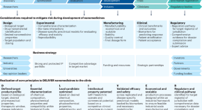
A translational framework to DELIVER nanomedicines to the clinic
The authors propose a framework to be followed during preclinical investigation of nanomedicines to increase their translatability potential.
- Christine J. Allen
- Hélder A. Santos
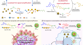
Self-immolative poly(thiocarbamate) with localized H 2 S signal amplification for precise cancer imaging and therapy
Hydrogen sulfide is essential in many biological processes and a promising cancer imaging and signalling molecule and therapeutic agent, but the potential applications are hindered by its low endogenous levels. Here, the authors develop a nanoplatform based on H 2 S-responsive self-immolative poly(thiocarbamate) with localized H 2 S signal amplification capability and use the nanoplatform to encapsulate an H 2 S-responsive fluorescent probe or an anticancer prodrug.
- Qingyu Zong
- Youyong Yuan
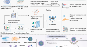
AI-powered omics-based drug pair discovery for pyroptosis therapy targeting triple-negative breast cancer
Cancer-targeted drug discovery can be achieved by transcriptomics screening on patients. Here this group reports a drug target screening model built upon triple-negative breast cancer (TNBC) cohort and drug database with the selected drug pair exhibiting effective pyroptosis induction and TNBC tumor growth inhibition.
- Boshu Ouyang
- Caihua Shan
- Zhiqing Pang
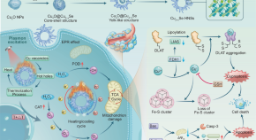
A singular plasmonic-thermoelectric hollow nanostructure inducing apoptosis and cuproptosis for catalytic cancer therapy
Thermoelectric catalytic therapy is an emerging therapeutic approach but faces the issue of limited temperature variations in living organisms. Here the authors address this issue by developing urchin-like Cu 2−x Se hollow nanospheres that display a cascade of plasmonic photothermal and thermoelectric conversion processes for plasmonic-thermoelectric catalytic cancer therapy.
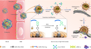
Polymer-locking fusogenic liposomes for glioblastoma-targeted siRNA delivery and CRISPR–Cas gene editing
Delivering gene editing materials to the brain for glioblastoma therapy can boost the efficacy of chemotherapy. Here the authors reduce resistance to temozolomide using a reactive oxygen species-sensitive polymer-locking fusogenic liposome that can cross the blood–brain barrier and deliver short interfering RNA or CRISPR–Cas to glioblastoma with high specificity.
- Jinquan Cai
News and Comment

Microrobots neutralize proinflammatory cytokines in the gut
An article in Science Robotics reports green algae-based microrobots, carrying macrophage membrane-coated nanoparticles, that can be orally administered to neutralize proinflammatory cytokines in the gastrointestinal tract to treat inflammatory bowel disease.
- Christine-Maria Horejs

The power of putting education first
From high school to distinguished professor of chemistry at Rhodes University, Tebello Nyokong discusses her inspiration and ambitions to promote science in South Africa.
- Tebello Nyokong
- Stephanie Greed
Chemotherapy delivery activated by radiation
An article in Advanced Materials presents a drug delivery platform for cancer chemotherapy that is activated by radiation.
- Charlotte Allard
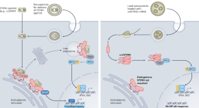
Controlling the STING pathway to improve immunotherapy
A genetically engineered variant of the stimulator of interferon genes (STING) protein is delivered to cancer cells, showing potential for clinical impact.
- John T. Wilson
Quick links
- Explore articles by subject
- Guide to authors
- Editorial policies

IMAGES
VIDEO
COMMENTS
Dental Biomaterials: Addressing Modern Challenges and Shaping Future Procedures. An interdisciplinary journal across nanoscience and nanotechnology, at the interface of chemistry, physics, materials science and engineering. It focuses on new nanofabrication methods and their ap...
Read the latest Research articles from Nature Nanotechnology. ... Nature Nanotechnology (Nat. Nanotechnol.) ISSN 1748-3395 (online) ISSN 1748-3387 (print) nature.com sitemap ...
RSS Feed. Nanoscience and technology is the branch of science that studies systems and manipulates matter on atomic, molecular and supramolecular scales (the nanometre scale). On such a length ...
Selected Topics in Nanoscience and Nanotechnology contains a collection of papers in the subfields of scanning probe microscopy, nanofabrication, functional nanoparticles and nanomaterials, molecular engineering and bionanotechnology. Written by experts in their respective fields, it is intended for a general scientific readership who may be non-specialists in these subjects, but who want a ...
Nanotechnology is one of the most promising key enabling technologies of the 21st century. The field of nanotechnology was foretold in Richard Feynman's famous 1959 lecture "There's Plenty of Room at the Bottom", and the term was formally defined in 1974 by Norio Taniguchi. Thus, the field is now approaching 50 years of research and application. It is a continuously expanding area of ...
Nature Nanotechnology offers a unique mix of news and reviews alongside top-quality research papers. Published monthly, in print and online, the journal reflects the entire spectrum of ...
Nanotechnology is a relatively new field of science and technology that studies tiny objects (0.1-100 nm). Due to various positive attributes displayed by the biogenic synthesis of nanoparticles (NPs) such as cost-effectiveness, none to negligible environmental hazards, and biological reduction served as an attractive alternative to its counterpart chemical methods.
Nanoscience teaching and research program in South Africa. Robert Lindsay. Janske Nel. Frontiers in Nanotechnology. doi 10.3389/fnano.2024.1401598. 1,258 views. An interdisciplinary journal across nanoscience and nanotechnology, at the interface of chemistry, physics, materials science and engineering. It focuses on new nanofabrication methods ...
Journal of Nanotechnology is an open access journal that publishes papers related to the science and technology of nanosized and nanostructured materials, with emphasis on their design, characterization, functionality, and preparation for implementation in systems and devices. As part of Wiley's Forward Series, this journal offers a ...
Abstract. Nanotechnology, contrary to its name, has massively revolutionized industries around the world. This paper predominantly deals with data regarding the applications of nanotechnology in the modernization of several industries. A comprehensive research strategy is adopted to incorporate the latest data driven from major science platforms.
Between 2007 and 2011, approximately € 896 million was invested by the EU alone in nanotechnology-related research. The investment in nanotechnology worldwide is estimated to be close to a quarter of a trillion USD, with both China and The USA investing upwards of US$ 2 billion. 9 While these two countries are considered nanotechnology giants ...
With software facilitated data mining approach, the journal scope and research topic of interest of Nano Today are analyzed.. Biomedicine and energy production/storage are identified as hot research areas in Nano Today.. Nano Today is featured as a multidisciplinary journal.. Primary research papers with time-sensitive topic in nanoscience and nanotechnology become a driving force for Nano Today.
There was a progress in nanotechnology since the early ideas of Feynman until 1981 when the physicists Gerd Binnig and Heinrich Rohrer invented a new type of microscope at IBM Zurich Research Laboratory, the Scanning Tunneling Microscope (STM) [19,20]. The STM uses a sharp tip that moves so close to a conductive surface that the electron wave ...
Nanodentistry is a separate branch of nanomedicine that involves a broad range of applications of nanotechnology ranging from detection to diagnosis, to cure treatment options and prognostic details about tooth functions [99]. A wide spectrum of oral health-related issues can be dealt with using nanomaterials [100].
Nanotechnology can be utilised for medication to particular cells in the body, thereby reducing the risks of failure and rejection. 17, 18, 19 We have identified four primary research objectives of this paper as under: (1) to identify types of Nanotechnology and Nanoparticles with their uses in the medical field; (2) to discuss classes and ...
A light-fuelled nanoratchet shifts a coupled chemical equilibrium. An artificial molecular machine was designed by coupling a chemical equilibrium to a photoresponsive molecular motor. Upon light ...
In this context, the aim of this Special Issue, entitled " Nanotechnology for Electronic Materials and Devices ", is to collect dedicated papers in several nanotechnological fields. The issue consists of eleven selected regular papers focusing on the latest developments in nanomaterials and nanotechnologies for electronic devices and sensors.
Nanotechnology is the study of the controlling the matter on an atom and molecular scale. Generally nanotechnology deals with structures sized between 1-100 nanometers in at least one. dimension ...
Research Open Access 12 Sept 2024 Microsystems & Nanoengineering. Volume: 10, P: 127. ... News & Views 15 Jul 2024 Nature Nanotechnology. P: 1-2. A light-touch approach to intracellular delivery.
1. Introduction. Nanotechnology and nanodelivery systems are innovative areas of science that comprise the design, characterization, manufacturing, and application of materials, devices, and systems at the nanoscale level (1-100 nm). Nanotechnology, being recognized as one of the revolutionizing technologies, is extensively studied in the ...
Over the last decade, increased knowledge of the microenvironment of tumors has motivated our efforts to develop nanoparticles as a novel cancer-related therapeutic and diagnostic strategy 2. Cancer tissues consist of non-cellular (e.g. interstitial and vascular) or cellular compartments which vary considerably from the healthy tissues around them.
Nanomedicine is a branch of medicine that applies the knowledge and tools of nanotechnology to the prevention and treatment of disease. Nanomedicine involves the use of nanoscale materials, such ...
Nanotechnology has made a huge impact on science, technology, and society. At the same time, educational institutions at all levels have recognized its relevance and introduced courses that aim at familiarizing their students with the basic concepts of nanotechnology. While the importance of teaching nanotechnology, the contents of such courses, and insightful activities in the laboratory have ...